추천 제품
생물학적 소스
rabbit
Quality Level
항체 형태
affinity isolated antibody
항체 생산 유형
primary antibodies
클론
polyclonal
정제법
affinity chromatography
종 반응성(상동성에 의해 예측)
all
제조업체/상표
Chemicon®
기술
ELISA: suitable
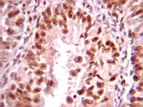
immunohistochemistry: suitable
immunoprecipitation (IP): suitable
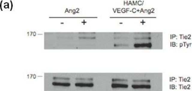
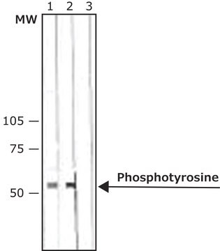
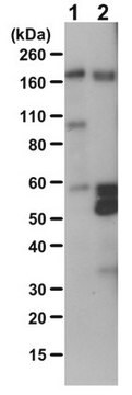
western blot: suitable
배송 상태
wet ice
타겟 번역 후 변형
phosphorylation (pSer)
일반 설명
Cellular responses to a variety of extracellular signals occur through phosphorylation or dephosphorylation of intracellular proteins. Abnormal protein phosphorylation may be involved in the progression of numerous diseases, including many forms of cancer, immune system dysfunction and cardiovascular disease. Such changes in protein phosphorylation may be detected by immunocytochemical mapping of cells labeled with anti-phosphoserine or anti-phosphothreonine antibodies and can be correlated with cellular, electrophysiological or behavioral state changes.
특이성
Anti-phosphoserine is species independent.
Recognizes phosphoserine, peptidyl-phosphoserine, and serine-phosphorylated proteins. Does not cross react with ATP, phosphotyrosine, peptidyl phosphothreonine and serine. Slight cross reactivity with free phosphothreonine. Readily reacts with known phosphoproteins such as phosvitin and alpha casein. Anti-phosphoserine is species independent in reactivity
면역원
Keyhole Limpet Hemocyanin(KLH)- conjugated phosphoserine conjugates.
애플리케이션
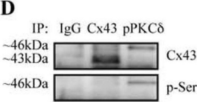
Anti-Phosphoserine Antibody detects level of Phosphoserine & has been published & validated for use in ELISA, IH, IP & WB.
Immunoprecipitation (tissue extracts):
10-20 μg anti-pS/mg protein was used from a previous lot.
Note that this will immunoprecipitate all phosphoserine proteins in the extracts. If using purified protein exacts containing only the protein of interest, antibody amounts should be decreased to 5 μg/500 μL of extracts. Optimal concentrations must be determined experimentally.
ELISA (kinase assay):
A previous lot of this anitbody was used at a 1:250-1:500 dilution.
Immunohistochemistry:
A previous lot of this antibody was used at a 1:50 dilution.
As the antibody detects all phosphoserines, any phosphorylated serine protein or peptide can be used to block antibody staining.
Typically a 10 M excess of peptide is used in Tris based buffers.
Western Blot Analysis:
1:500 dilution of a previous lot
Optimal working dilutions must be determined by end user.
Research Category
Signaling
Signaling
Research Sub Category
General Post-translation Modification
General Post-translation Modification
품질
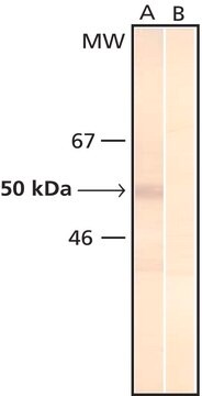
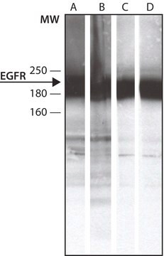
Routinely evaluated by Western Blot on NGF treated PC12 lysates.
Western Blot Analysis: 1:500 dilution of this lot detected serine phosphorylated proteins on 10 μg of NGF treated PC12 lysates.
Western Blot Analysis: 1:500 dilution of this lot detected serine phosphorylated proteins on 10 μg of NGF treated PC12 lysates.
표적 설명
Dependent upon the molecular weight of the serine phosphorylated protein being detected.
물리적 형태
Phosphoserine affinity chromatography
Purified rabbit serum in buffer containing 50% glycerol.
저장 및 안정성
Stable for up to 6 months at -20°C in undiluted aliquots from date of receipt. During shipment, small volumes of product will occasionally become entrapped in the seal of the product vial. For products with volumes of 200 uL or less, we recommend gently tapping the vial on a hard surface or briefly centrifuging the vial in a tabletop centrifuge to dislodge any liquid in the container′s cap.
분석 메모
Control
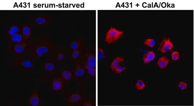
NIH 3T3 cells (+/- TPA), K562 cells, and EGF-stimulated A431 cells.
NIH 3T3 cells (+/- TPA), K562 cells, and EGF-stimulated A431 cells.
기타 정보
Concentration: Please refer to the Certificate of Analysis for the lot-specific concentration.
법적 정보
CHEMICON is a registered trademark of Merck KGaA, Darmstadt, Germany
면책조항
Unless otherwise stated in our catalog or other company documentation accompanying the product(s), our products are intended for research use only and are not to be used for any other purpose, which includes but is not limited to, unauthorized commercial uses, in vitro diagnostic uses, ex vivo or in vivo therapeutic uses or any type of consumption or application to humans or animals.
적합한 제품을 찾을 수 없으신가요?
당사의 제품 선택기 도구.을(를) 시도해 보세요.
Storage Class Code
12 - Non Combustible Liquids
WGK
WGK 2
Flash Point (°F)
Not applicable
Flash Point (°C)
Not applicable
시험 성적서(COA)
제품의 로트/배치 번호를 입력하여 시험 성적서(COA)을 검색하십시오. 로트 및 배치 번호는 제품 라벨에 있는 ‘로트’ 또는 ‘배치’라는 용어 뒤에서 찾을 수 있습니다.
이미 열람한 고객
Short-term in vivo inhibition of insulin receptor substrate-1 expression leads to insulin resistance, hyperinsulinemia, and increased adiposity.
Araujo, EP; De Souza, CT; Gasparetti, AL; Ueno, M; Boschero, AC; Saad, MJ; Velloso, LA
Endocrinology null
Activated NAD(P)H oxidase from supplemental oxygen induces neovascularization independent of VEGF in retinopathy of prematurity model.
Saito, Y; Uppal, A; Byfield, G; Budd, S; Hartnett, ME
Investigative Ophthalmology & Visual Science null
Protein kinase C-mediated modulation of FIH-1 expression by the homeodomain protein CDP/Cut/Cux.
Li, J; Wang, E; Dutta, S; Lau, JS; Jiang, SW; Datta, K; Mukhopadhyay, D
Molecular and cellular biology null
Pkd2+/- vascular smooth muscles develop exaggerated vasocontraction in response to phenylephrine stimulation.
Qian, Q; Hunter, LW; Du, H; Ren, Q; Han, Y; Sieck, GC
Journal of the American Society of Nephrology null
A novel high throughput biochemical assay to evaluate the HuR protein-RNA complex formation.
D'Agostino, VG; Adami, V; Provenzani, A
Testing null
자사의 과학자팀은 생명 과학, 재료 과학, 화학 합성, 크로마토그래피, 분석 및 기타 많은 영역을 포함한 모든 과학 분야에 경험이 있습니다..
고객지원팀으로 연락바랍니다.