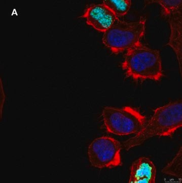
04-889
Anti-Src Antibody, clone N6L, rabbit monoclonal
culture supernatant, clone N6L, from rabbit
동의어(들):
proto-oncogene tyrosine-protein kinase SRC, protooncogene SRC, Rous sarcoma, tyrosine kinase pp60c-src, tyrosine-protein kinase SRC-1, v-src avian sarcoma (Schmidt-Ruppin A-2) viral oncogene
homolog, v-src sarcoma (Schmidt-Ruppin A-2) viral onco
로그인조직 및 계약 가격 보기
모든 사진(1)
About This Item
UNSPSC 코드:
12352203
eCl@ss:
32160702
NACRES:
NA.41
추천 제품
생물학적 소스
rabbit
Quality Level
항체 형태
culture supernatant
항체 생산 유형
primary antibodies
클론
N6L, monoclonal
종 반응성
human
기술
western blot: suitable
동형
IgG
NCBI 수납 번호
UniProt 수납 번호
배송 상태
dry ice
타겟 번역 후 변형
unmodified
유전자 정보
human ... SRC(6714)
일반 설명
pp60c-Src is the prototype non-receptor tyrosine kinase. Homology to Src in three regions is shared by a number of proteins: SH1 being the catalytic domain, SH2 being a phospho-tyrosine binding domain, and SH3 being a proline-binding domain. The Src SH2 domain binds to phospho-Tyr527 (phosphorylated by Csk) within the Src molecule, causing Src to adopt an inactive conformation. Mutation of Tyr527 to phenylalanine removes the SH2-binding target and results in a constitutively active kinase. A second mutation at the conserved lysine residue at the ATP-binding site abolishes kinase activity, but without Tyr527, the conformation of the protein is open, allowing interaction with endogenous activators. Consequently, this double-mutant exhibits a dominant-negative phenotype
특이성
Recognizes Src. Crossreacts with Yes and weakly with Fyn. No crossreactivity observed with Lck or Lyn.
면역원
N-terminal 6His-tagged recombinant full-length human Src expressed by baculovirus in Sf21 insect cells. Purified using Ni2+/NTA agarose.
애플리케이션
Research Category
Cell Structure
Cell Structure
Research Sub Category
Cytoskeletal Signaling
Cytoskeletal Signaling
This Anti-Src Antibody, clone N6L is validated for use in WB for the detection of Src.
품질
Western Blot Analysis: A 1:500 to 1:1000 dilution of this antibody detected Src in RIPA lysates from A431 cells.
표적 설명
~ 60 kDa
결합
Replaces: 05-889
물리적 형태
Cultured supernantant containing 0.05% sodium azide.
저장 및 안정성
Stable for 1 year at -20ºC from date of receipt.
Handling Recommendations: Upon receipt, and prior to removing the cap, centrifuge the vial and gently mix the solution. Aliquot into microcentrifuge tubes and store at -20°C. Avoid repeated freeze/thaw cycles, which may damage IgG and affect product performance.
Handling Recommendations: Upon receipt, and prior to removing the cap, centrifuge the vial and gently mix the solution. Aliquot into microcentrifuge tubes and store at -20°C. Avoid repeated freeze/thaw cycles, which may damage IgG and affect product performance.
분석 메모
Control
Included positive antigen control of non-stimulated A431 lysate (Cat. No. 12-301). Add 2.5 μL of 2-mercaptoethanol/100 μL of lysate and boil for 5 minutes to reduce the preparation. Load 20 μg of reduced lysate per lane for minigels.
Included positive antigen control of non-stimulated A431 lysate (Cat. No. 12-301). Add 2.5 μL of 2-mercaptoethanol/100 μL of lysate and boil for 5 minutes to reduce the preparation. Load 20 μg of reduced lysate per lane for minigels.
기타 정보
Concentration: Please refer to the Certificate of Analysis for the lot-specific concentration.
면책조항
Unless otherwise stated in our catalog or other company documentation accompanying the product(s), our products are intended for research use only and are not to be used for any other purpose, which includes but is not limited to, unauthorized commercial uses, in vitro diagnostic uses, ex vivo or in vivo therapeutic uses or any type of consumption or application to humans or animals.
적합한 제품을 찾을 수 없으신가요?
당사의 제품 선택기 도구.을(를) 시도해 보세요.
Storage Class Code
12 - Non Combustible Liquids
WGK
WGK 1
Flash Point (°F)
Not applicable
Flash Point (°C)
Not applicable
시험 성적서(COA)
제품의 로트/배치 번호를 입력하여 시험 성적서(COA)을 검색하십시오. 로트 및 배치 번호는 제품 라벨에 있는 ‘로트’ 또는 ‘배치’라는 용어 뒤에서 찾을 수 있습니다.
Xiang Li et al.
Kardiologia polska, 79(9), 972-979 (2021-06-28)
Interleukin (IL)-18 is produced mainly in the heart and can be associated with the development of cardiac hypertrophy that leads to cardiac dysfunction. However, the effects of hypoxia on IL-18 expression and atrial natriuretic factor (ANF) secretion remain largely unknown.
Mariya S Liyasova et al.
PloS one, 14(5), e0216967-e0216967 (2019-05-24)
Many receptor tyrosine kinases (RTKs, such as EGFR, MET) are negatively regulated by ubiquitination and degradation mediated by Cbl proteins, a family of RING finger (RF) ubiquitin ligases (E3s). Loss of Cbl protein function is associated with malignant transformation driven
자사의 과학자팀은 생명 과학, 재료 과학, 화학 합성, 크로마토그래피, 분석 및 기타 많은 영역을 포함한 모든 과학 분야에 경험이 있습니다..
고객지원팀으로 연락바랍니다.