Skip to Content
Merck
All Photos(3)

Key Documents

SAB4200719

Sigma-Aldrich

Anti-Laminin antibody, Mouse monoclonal

enhanced validation

clone LAM-89, purified from hybridoma cell culture

Synonym(s):

Anti-LAMA, Anti-LAMA1

Sign Into View Organizational & Contract Pricing


About This Item

UNSPSC Code:
12352203
NACRES:
NA.41

biological source

mouse

Quality Level

antibody form

purified from hybridoma cell culture

antibody product type

primary antibodies

clone

LAM-89, monoclonal

form

buffered aqueous solution

mol wt

~850 kDa

species reactivity

feline, human, porcine

enhanced validation

independent
Learn more about Antibody Enhanced Validation

concentration

~1.0 mg/mL

technique(s)

ELISA: suitable
electron microscopy: suitable
immunoblotting: 0.03-0.06 μg/mL using Laminin from Engelbreth-Holm-Swarm murine sarcoma basement membrane
immunofluorescence: suitable
immunohistochemistry: 20-40 μg/mL using pronase-retrieved formalin-fixed, paraffin-embedded human tongue sections.

isotype

IgG1

UniProt accession no.

shipped in

dry ice

storage temp.

−20°C

target post-translational modification

unmodified

Gene Information

General description

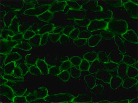
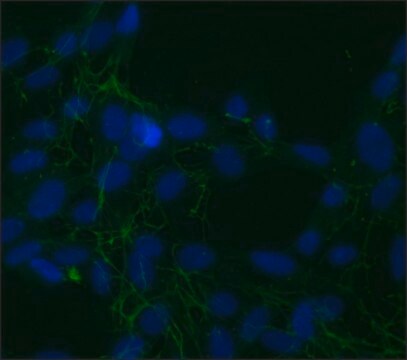
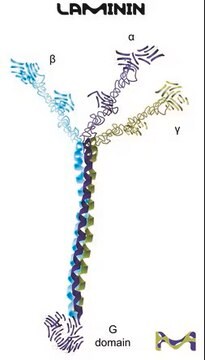
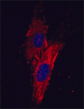
Monoclonal anti-laminin antibody (mouse IgG1 isotype) is derived from the LAM-89 hybridoma produced by the fusion of mouse myeloma cells and splenocytes from a mouse immunized with purified laminin from human placenta. Monoclonal anti-laminin antibody specifically recognizes laminin from human, feline and porcine origin. The antibody shows reactivity with laminin and specifically staining basal membrane of blood vessels, epithelium, nerve and muscle fiber. Monoclonal anti-laminin shows no reaction with collagen IV, fibronectin, vitronectin and chondroitin sulfate type A, B or C. Laminins are complexed extracellular glycoproteins which assemble the basal components of the basement membrane (BM). Each laminin is a heterotrimer (~850kDa) comprising of an α (~400kDa), β (~200kDa) and γ (~200kDa) chains all held together by disulfide bonds.

Specificity

Monoclonal Anti-Laminin antibody specifically recognizes Laminin from human, feline and porcine origin.

Immunogen

purified Laminin from human Placenta

Application

Monoclonal Anti-Laminin is recommended to use in various immunochemical assays, including immunohistochemistry, immunoblotting (~850 kDa), immunofluorescence, Enzyme linked immunosorbent assay (ELISA) and electron microscopy.

Biochem/physiol Actions

Laminins contribute to the structure of the extracellular matrix (ECM). It also plays a major role in the regulation of variety of cellular processes, such as the rates of cell proliferation, differentiation, adhesion, migration, cell shaping, phenotype stability and resistance to apoptosis. Laminin polymers forms supramolecular network with collagen IV polymers through heparan sulfate proteoglycan and by nidogen (entactin) and plays an important role in the basement membrane (BM) stability. In mouse, mutations in the gene is associated with the development of muscular dystrophy.

Physical form

Solution in 0.01 M phosphate buffered saline, pH 7.4, containing 15 mM sodium azide.

Storage and Stability

Store at –20°C. For continuous use, store at 2–8°C for up to one month. For extended storage, freeze in working aliquots. Repeated freezing and thawing is not recommended. If slight turbidity occurs upon prolonged storage, clarify the solution by centrifugation before use. Working dilution samples should be discarded if not used within 12 hours.

Disclaimer

Unless otherwise stated in our catalog or other company documentation accompanying the product(s), our products are intended for research use only and are not to be used for any other purpose, which includes but is not limited to, unauthorized commercial uses, in vitro diagnostic uses, ex vivo or in vivo therapeutic uses or any type of consumption or application to humans or animals.

Not finding the right product?  

Try our Product Selector Tool.

Storage Class Code

10 - Combustible liquids

WGK

WGK 1

Flash Point(F)

Not applicable

Flash Point(C)

Not applicable


Choose from one of the most recent versions:

Certificates of Analysis (COA)

Lot/Batch Number

Don't see the Right Version?

If you require a particular version, you can look up a specific certificate by the Lot or Batch number.

Already Own This Product?

Find documentation for the products that you have recently purchased in the Document Library.

Visit the Document Library

Laminins: Roles and Utility in Wound Repair.
Iorio V, et al.
Advances in Wound Care, 4, 250-263 (2015)
Murine muscular dystrophy caused by a mutation in the laminin alpha 2 (Lama2) gene.
Xu H, et al.
Nature Genetics, 8, 297-302 (1994)
Basic components of connective tissues and extracellular matrix: elastin, fibrillin, fibulins, fibrinogen, fibronectin, laminin, tenascins and thrombospondins.
Halper J and Kjaer M
Advances in Experimental Medicine and Biology, 31-47 (2014)
The laminin family
Aumailley M, et al.
Cell adhesion & migration, 7, 48-55 (2013)
Junya Takegaki et al.
Scientific reports, 11(1), 21224-21224 (2021-10-29)
Skeletal muscle mass is critical for good quality of life. Mesenchymal stem cells (MSCs) are multipotent stem cells distributed across various tissues. They are characterized by the capacity to secrete growth factors and differentiate into skeletal muscle cells. These capabilities

Our team of scientists has experience in all areas of research including Life Science, Material Science, Chemical Synthesis, Chromatography, Analytical and many others.

Contact Technical Service