11922416001
Roche
Anti-His6
from mouse IgG1
Synonym(s):
metal chelate affinity chromatography
Sign Into View Organizational & Contract Pricing
All Photos(1)
About This Item
UNSPSC Code:
12352203
Recommended Products
biological source
mouse
Quality Level
conjugate
unconjugated
antibody form
purified immunoglobulin
antibody product type
primary antibodies
clone
monoclonal
form
lyophilized
packaging
pkg of 100 μg
manufacturer/tradename
Roche
isotype
IgG1
storage temp.
2-8°C
General description
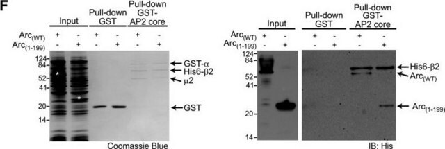
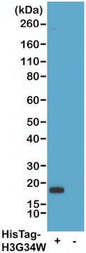
Anti-His6 is a monoclonal antibody to His6-tagged proteins. Anti-His6 specifically recognizes an epitope of six consecutive histidine residues of both natural and recombinant proteins. The antibody reacts with native and denatured histidine-tagged fusion proteins independent of the location of the histidine-tag in the protein sequence.
Specificity
Anti-His6 specifically recognizes an epitope of six consecutive histidine residues (His6) in natural and recombinant proteins. It reacts with both native and denatured histidine-tagged fusion proteins, independent of location of the histidine-tag in the protein sequence. In addition, Anti-His6 preferentially recognizes the C-terminal His6-epitope with high sensitivity. Anti-His6 allows specific and sensitive detection of histidine-tagged proteins irrespective of the expression system used.
The antibody recognizes an epitope of six consecutive histidine residues (His6) in natural and recombinant proteins. It reacts with both native and denatured histidine-tagged fusion proteins, independent of the epitope-sequence location.
Application
Anti-His6 is used for the detection of His6-tagged proteins in:
- Immunoblotting (e.g., dot blots and western blots)
- Immunoprecipitation
- Immunoassays (ELISA)
- Immunocytochemistry
- Immunofluorescence
Preparation Note
Stabilizers: BSA
Working concentration: Working concentration of conjugate depends on application and substrate.
The following concentrations should be taken as a guideline:
Working concentration: Working concentration of conjugate depends on application and substrate.
The following concentrations should be taken as a guideline:
- ELISA: for detection 0.1 μg/ml; for coating 1 to 5 μg/ml
- Immunoprecipitation: 0.5 to 5 μg/ml
- Western blot: 0.2 to 0.5 μg/ml
Reconstitution
Add 1 ml double-distilled water to a final concentration of 100 μg/ml.
Other Notes
For life science research only. Not for use in diagnostic procedures.
Not finding the right product?
Try our Product Selector Tool.
Signal Word
Warning
Hazard Statements
Precautionary Statements
Hazard Classifications
Aquatic Chronic 3 - Skin Sens. 1
Storage Class Code
11 - Combustible Solids
WGK
WGK 2
Flash Point(F)
does not flash
Flash Point(C)
does not flash
Choose from one of the most recent versions:
Already Own This Product?
Find documentation for the products that you have recently purchased in the Document Library.
Customers Also Viewed
Aniek D van der Woude et al.
Microbial cell factories, 15, 60-60 (2016-04-10)
Erythritol is a polyol that is used in the food and beverage industry. Due to its non-caloric and non-cariogenic properties, the popularity of this sweetener is increasing. Large scale production of erythritol is currently based on conversion of glucose by
Michael Kisiela et al.
The FEBS journal, 285(2), 275-293 (2017-11-21)
The human dehydrogenase/reductase SDR family member 4 (DHRS4) is a tetrameric protein that is involved in the metabolism of several aromatic carbonyl compounds, steroids, and bile acids. The only invertebrate DHRS4 that has been characterized to date is that from
Chandrika Canugovi et al.
DNA repair, 9(10), 1080-1089 (2010-08-27)
Mitochondrial transcription factor A (TFAM) is an essential component of mitochondrial nucleoids. TFAM plays an important role in mitochondrial transcription and replication. TFAM has been previously reported to inhibit nucleotide excision repair (NER) in vitro but NER has not yet
Donghyuk Shin et al.
Molecular cell, 77(1), 164-179 (2019-11-17)
The family of bacterial SidE enzymes catalyzes non-canonical phosphoribosyl-linked (PR) serine ubiquitination and promotes infectivity of Legionella pneumophila. Here, we describe identification of two bacterial effectors that reverse PR ubiquitination and are thus named deubiquitinases for PR ubiquitination (DUPs; DupA
Archana Kumari et al.
The EMBO journal, 39(14), e104006-e104006 (2020-06-23)
Cellular studies of filamentous actin (F-actin) processes commonly utilize fluorescent versions of toxins, peptides, and proteins that bind actin. While the choice of these markers has been largely based on availability and ease, there is a severe dearth of structural
Our team of scientists has experience in all areas of research including Life Science, Material Science, Chemical Synthesis, Chromatography, Analytical and many others.
Contact Technical Service


