MABC32
Anti-p62 (Sequestosome-1) Antibody, clone 11C9.2
clone 11C9.2, from mouse
Synonym(s):
sequestosome 1, EBI3-associated protein of 60 kDa, Paget disease of bone 3, phosphotyrosine independent ligand for the Lck SH2 domain p62, oxidative stress induced like, EBI3-associated protein p60, Phosphotyrosine-independent ligand for the Lck SH2 doma
About This Item
Recommended Products
biological source
mouse
Quality Level
antibody form
purified antibody
antibody product type
primary antibodies
clone
11C9.2, monoclonal
species reactivity
mouse, rat, human
technique(s)
flow cytometry: suitable
immunocytochemistry: suitable
western blot: suitable
isotype
IgMκ
NCBI accession no.
UniProt accession no.
shipped in
wet ice
target post-translational modification
unmodified
Gene Information
human ... SQSTM1(8878)
Related Categories
General description
Immunogen
Application
Apoptosis & Cancer
Apoptosis - Additional
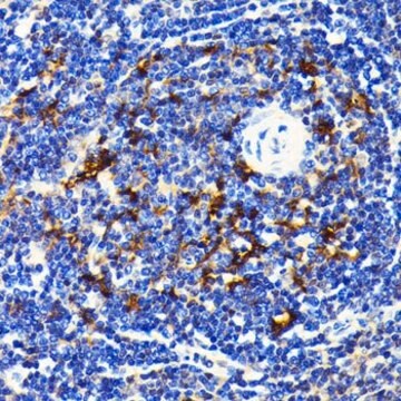
Flow Cytometry Analysis: 1.0 µg from a previous lot detected p62 (Sequestosome-1) in the staining of fixed and permeabilized HeLa cells.
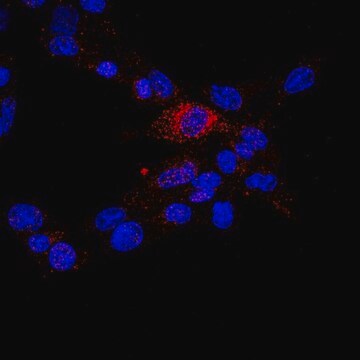
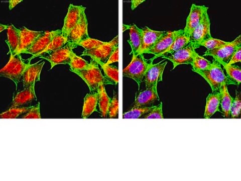
Immunocytochemistry Analysis: A 1:500 dilution from a previous lot detected p62 (Sequestosome-1) in NIH/3T3, A431, and HeLa cells.
Quality
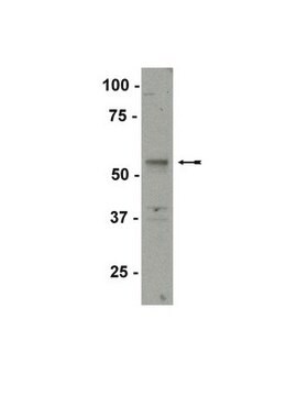
Western Blot Analysis: 0.001 µg/mL of this antibody detected p62 (Sequestosome-1) in 10 µg of A431 cell lysate.
Target description
The calculated molecular weight of this protein is 47 kDa and also has an isoform at 38 kDa Due to modifications, this protein may be observed up to ~60 kDa in some lysates.
Physical form
Storage and Stability
Analysis Note
A431 cell lysate
Disclaimer
Not finding the right product?
Try our Product Selector Tool.
recommended
Storage Class Code
12 - Non Combustible Liquids
WGK
WGK 2
Flash Point(F)
Not applicable
Flash Point(C)
Not applicable
Certificates of Analysis (COA)
Search for Certificates of Analysis (COA) by entering the products Lot/Batch Number. Lot and Batch Numbers can be found on a product’s label following the words ‘Lot’ or ‘Batch’.
Already Own This Product?
Find documentation for the products that you have recently purchased in the Document Library.
Articles
Autophagy is a highly regulated process that is involved in cell growth, development, and death. In autophagy cells destroy their own cytoplasmic components in a very systematic manner and recycle them.
Our team of scientists has experience in all areas of research including Life Science, Material Science, Chemical Synthesis, Chromatography, Analytical and many others.
Contact Technical Service

