Skip to Content
Merck
All Photos(2)

Key Documents

Safety Information

SAB4500362

Sigma-Aldrich

Anti-Collagen I antibody produced in rabbit

affinity isolated antibody

Synonym(s):

α 2 type i collagen, co1a2, col1a2, collagen, type i

Sign Into View Organizational & Contract Pricing


About This Item

UNSPSC Code:
12352203
NACRES:
NA.41

biological source

rabbit

Quality Level

conjugate

unconjugated

antibody form

affinity isolated antibody

antibody product type

primary antibodies

clone

polyclonal

form

buffered aqueous solution

mol wt

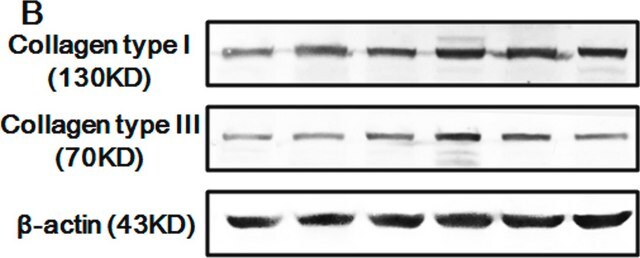
antigen 129 kDa

species reactivity

mouse, human, rat

concentration

~1 mg/mL

technique(s)

ELISA: 1:5000
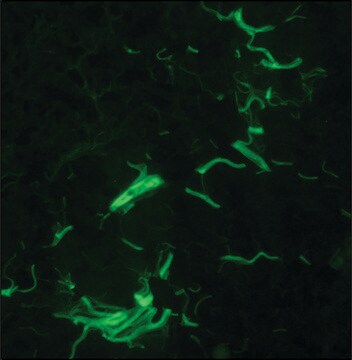
immunofluorescence: 1:100-1:500
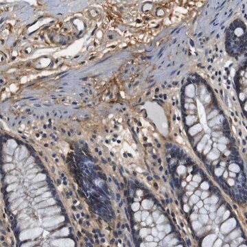
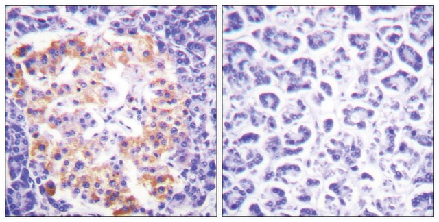
immunohistochemistry: 1:50-1:100

NCBI accession no.

UniProt accession no.

shipped in

wet ice

storage temp.

−20°C

target post-translational modification

unmodified

Gene Information

human ... COL1A2(1278)

General description

Collagen I is encoded by the gene COL1A2 (collagen type I alpha 2). The gene is located on human chromosome 7q21.3. COL1A2 is a fibril forming collagen, found in bone, dermis, tendon, ligaments and cornea.
Collagen is the major structural component of extracellular matrices found in connective tissues and internal organs, but is most prevalent in the dermis, tendons, and bones. It is classified into a number of structurally and genetically distinct types. Type I collagen is a heterodimer composed of two α1(I) chains and one α2(I) chain that spontaneously forms a triple helix scaffold at neutral pH and 37 °C. Collagen type I is an excellent substrate for the culture of hepatocytes, fibroblasts, spinal ganglion, muscle cells, Schwann cells, embryonic lung cells, epithelial cells, and a number of other cell lines. It has also been used in the study of growth, differentiation, migration of cell lines, and tissue morphogenesis during development.
Rabbit polyclonal anti-Collagen I, N-Terminal antibody reacts with mouse, rat, and human type I collagens.

Immunogen

The antiserum was produced against synthesized peptide derived from human Collagen I.

Immunogen Range: 1-50

Application

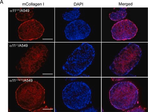
Anti-Collagen I antibody produced in rabbit has been used in western blotting and immunostaining.
Applications in which this antibody has been used successfully, and the associated peer-reviewed papers, are given below.
Immunofluorescence (1 paper)
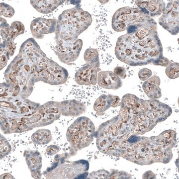
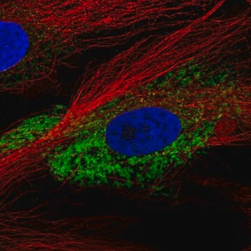
Rabbit polyclonal anti-Collagen I, N-Terminal antibody is used to tag collagen type I for detection and quantitation by immunocytochemical and immunohistochemical (IHC) techniques such as ELISA, immunoblotting, and immunoprecipitation. It is used as a probe to determine the presence and roles of collagen type I as a cell substrate.

Biochem/physiol Actions

Collagen I interacts with cytoskeleton and regulates cell motility. Mutations in this gene is linked to osteoporotic fracture, osteogenesis imperfecta and recessive classical type Ehlers-Danlos syndrome. It is also associated with idiopathic osteoporosis and atypical Marfan syndrome.

Features and Benefits

Evaluate our antibodies with complete peace of mind. If the antibody does not perform in your application, we will issue a full credit or replacement antibody. Learn more.

Physical form

Rabbit IgG in phosphate buffered saline (without Mg2+ and Ca2+), pH 7.4, 150mM NaCl, 0.02% sodium azide and 50% glycerol.

Disclaimer

Unless otherwise stated in our catalog or other company documentation accompanying the product(s), our products are intended for research use only and are not to be used for any other purpose, which includes but is not limited to, unauthorized commercial uses, in vitro diagnostic uses, ex vivo or in vivo therapeutic uses or any type of consumption or application to humans or animals.

Not finding the right product?  

Try our Product Selector Tool.

Storage Class Code

12 - Non Combustible Liquids

WGK

nwg

Flash Point(F)

Not applicable

Flash Point(C)

Not applicable


Regulatory Listings

Regulatory Listings are mainly provided for chemical products. Only limited information can be provided here for non-chemical products. No entry means none of the components are listed. It is the user’s obligation to ensure the safe and legal use of the product.

JAN Code

SAB4500362-100UG:


Certificates of Analysis (COA)

Search for Certificates of Analysis (COA) by entering the products Lot/Batch Number. Lot and Batch Numbers can be found on a product’s label following the words ‘Lot’ or ‘Batch’.

Already Own This Product?

Find documentation for the products that you have recently purchased in the Document Library.

Visit the Document Library

Respiratory syncytial virus infection accelerates lung fibrosis through the unfolded protein response in a bleomycin-induced pulmonary fibrosis animal model
Wang L, et al.
Molecular Medicine Reports, 16(1), 310-316 (2017)
Yangming Xiao et al.
Annals of biomedical engineering, 44(9), 2840-2850 (2016-02-26)
Artery buckling alters the fluid shear stress and wall stress in the artery but its temporal effect on vascular wall remodeling is poorly understood. The purpose of this study was to investigate the early effect of artery buckling on endothelial
COL1A2 is a Novel Biomarker to Improve Clinical Prediction in Human Gastric Cancer: Integrating Bioinformatics and Meta-Analysis
Rong L, et al.
Pathology Oncology Research, 24(1), 129-134 (2018)
Collagenase 1A2 (COL 1A2) gene A/C polymorphism in relation to severity of dental fluorosis
Escobar-Garcia D, et al.
Community dentistry and oral epidemiology, 44(2), 162-168 (2016)
Long-term characterization of axon regeneration and matrix changes using multiple channel bridges for spinal cord regeneration
Tuinstra HM, et al.
Tissue Engineering: Part A, 20(5-6), 1027-1037 (2013)

Our team of scientists has experience in all areas of research including Life Science, Material Science, Chemical Synthesis, Chromatography, Analytical and many others.

Contact Technical Service