Skip to Content
Merck
All Photos(9)

Documents

Safety Information

HPA049854

Sigma-Aldrich

Anti-PACSIN2 antibody produced in rabbit

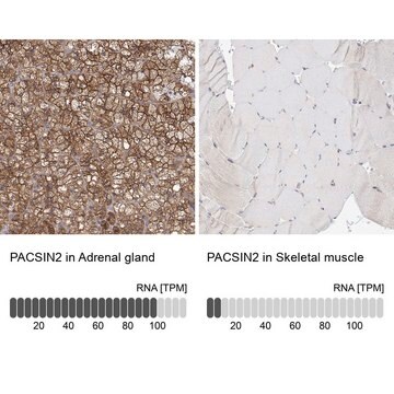
enhanced validation

Prestige Antibodies® Powered by Atlas Antibodies, affinity isolated antibody, buffered aqueous glycerol solution

Synonym(s):

Anti-SDPII, Anti-protein kinase C and casein kinase substrate in neurons 2

Sign Into View Organizational & Contract Pricing


About This Item

UNSPSC Code:
12352203
Human Protein Atlas Number:
NACRES:
NA.43

biological source

rabbit

conjugate

unconjugated

antibody form

affinity isolated antibody

antibody product type

primary antibodies

clone

polyclonal

product line

Prestige Antibodies® Powered by Atlas Antibodies

form

buffered aqueous glycerol solution

species reactivity

human

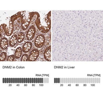
enhanced validation

independent
orthogonal RNAseq
Learn more about Antibody Enhanced Validation

technique(s)

immunoblotting: 0.04-0.4 μg/mL
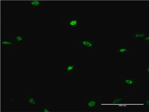
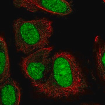
immunofluorescence: 0.25-2 μg/mL
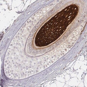
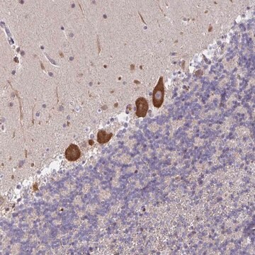
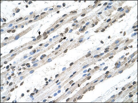
immunohistochemistry: 1:200-1:500

immunogen sequence

NVPSNPAQSAQSQSSYNPFEDEDDTGSTVSEKDDTKAKNVSSYEKTQSYPTDWS

UniProt accession no.

shipped in

wet ice

storage temp.

−20°C

target post-translational modification

unmodified

Gene Information

human ... PACSIN2(11252)

General description

PACSIN2 (protein kinase C and casein kinase substrate in neurons 2) is also known as syndapin-II. It has a Fer-CIP4 homology-BAR (F-BAR) domain. It is widely expressed. It is located on chromosome 22q13.2. PACSIN2 is present close to the pointed ends of microvillar actin bundles.

Immunogen

protein kinase C and casein kinase substrate in neurons 2 recombinant protein epitope signature tag (PrEST)

Application

Applications in which this antibody has been used successfully, and the associated peer-reviewed papers, are given below.
Immunofluorescence (1 paper)
Western Blotting (1 paper)

Biochem/physiol Actions

Anti-PACSIN2 has been used in western blotting and immunofluorescence.
PACSIN2 (protein kinase C and casein kinase substrate in neurons 2) participates in the formation of caveolae and endocytosis. It participates in the internalization of epidermal growth factor receptor (EGFR) and entry of cholera toxin B (CTxB).

Features and Benefits

Prestige Antibodies® are highly characterized and extensively validated antibodies with the added benefit of all available characterization data for each target being accessible via the Human Protein Atlas portal linked just below the product name at the top of this page. The uniqueness and low cross-reactivity of the Prestige Antibodies® to other proteins are due to a thorough selection of antigen regions, affinity purification, and stringent selection. Prestige antigen controls are available for every corresponding Prestige Antibody and can be found in the linkage section.

Every Prestige Antibody is tested in the following ways:
  • IHC tissue array of 44 normal human tissues and 20 of the most common cancer type tissues.
  • Protein array of 364 human recombinant protein fragments.

Linkage

Corresponding Antigen APREST85317

Physical form

Solution in phosphate buffered saline, pH 7.2, containing 40% glycerol and 0.02% sodium azide.

Legal Information

Prestige Antibodies is a registered trademark of Merck KGaA, Darmstadt, Germany

Disclaimer

Unless otherwise stated in our catalog or other company documentation accompanying the product(s), our products are intended for research use only and are not to be used for any other purpose, which includes but is not limited to, unauthorized commercial uses, in vitro diagnostic uses, ex vivo or in vivo therapeutic uses or any type of consumption or application to humans or animals.

Not finding the right product?  

Try our Product Selector Tool.

Storage Class Code

10 - Combustible liquids

WGK

WGK 1

Flash Point(F)

Not applicable

Flash Point(C)

Not applicable


Regulatory Listings

Regulatory Listings are mainly provided for chemical products. Only limited information can be provided here for non-chemical products. No entry means none of the components are listed. It is the user’s obligation to ensure the safe and legal use of the product.

JAN Code

HPA049854-100UL:


Certificates of Analysis (COA)

Search for Certificates of Analysis (COA) by entering the products Lot/Batch Number. Lot and Batch Numbers can be found on a product’s label following the words ‘Lot’ or ‘Batch’.

Already Own This Product?

Find documentation for the products that you have recently purchased in the Document Library.

Visit the Document Library

Hypomethylation and Genetic Instability in Monosomy Blastocysts May Contribute to Decreased Implantation Potential.
PLoS ONE, 11(7):e0159507 (2016)
Clostridium difficile Toxin A Undergoes Clathrin-Independent, PACSIN2-Dependent Endocytosis.
Chandrasekaran R, et al.
PLoS Pathogens, 12(12):e1006070 (2016)
Cordon bleu promotes the assembly of brush border microvilli.
Grega-Larson N E, et al.
Molecular Biology of the Cell, 26(21), 3803-3815 (2015)
Qiang Feng et al.
Journal of cell science, 130(15), 2491-2505 (2017-06-10)
Misplaced formation of microvilli to basolateral domains and intracellular inclusions in enterocytes are pathognomonic features in congenital enteropathy associated with mutation of the apical plasma membrane receptor syntaxin 3 (STX3). Although the demonstrated binding of Myo5b to the Rab8a and
Nathan E Grega-Larson et al.
Molecular biology of the cell, 26(21), 3803-3815 (2015-09-12)
Microvilli are actin-based protrusions found on the surface of diverse cell types, where they amplify membrane area and mediate interactions with the external environment. In the intestinal tract, these protrusions play central roles in nutrient absorption and host defense and

Our team of scientists has experience in all areas of research including Life Science, Material Science, Chemical Synthesis, Chromatography, Analytical and many others.

Contact Technical Service