Skip to Content
Merck
All Photos(1)

Key Documents

Safety Information

MABF317

Sigma-Aldrich

Anti-CD3ε (mouse) Antibody, PE, clone 145-2C11

clone 145-2C11, from hamster(Armenian), PE

Synonym(s):

T-cell surface glycoprotein CD3 epsilon chain, T-cell surface antigen T3/Leu-4 epsilon chain, CD3e

Sign Into View Organizational & Contract Pricing


About This Item

UNSPSC Code:
12352203
eCl@ss:
32160702
NACRES:
NA.41
conjugate:
PE
application:
FACS
IHC
IP
WB
activity assay
clone:
145-2C11, monoclonal
species reactivity:
mouse
citations:
1
technique(s):
activity assay: suitable
flow cytometry: suitable
immunohistochemistry: suitable
immunoprecipitation (IP): suitable
western blot: suitable

biological source

hamster (Armenian)

conjugate

PE

antibody form

affinity isolated antibody

antibody product type

primary antibodies

clone

145-2C11, monoclonal

species reactivity

mouse

technique(s)

activity assay: suitable
flow cytometry: suitable
immunohistochemistry: suitable
immunoprecipitation (IP): suitable
western blot: suitable

isotype

IgG

UniProt accession no.

shipped in

wet ice

target post-translational modification

unmodified

Gene Information

mouse ... Cd3E(12501)

General description

CD3, also known as T3, is a member of the Ig and the ITAM superfamilies. The CD3 complex is composed of six polypeptides with usually four different transmembrane CD3 chains, gamma, delta, epsilon and zeta. CD3 is primarily expressed on T cells, NK-T cells, and at different levels on thymocytes during T cell differentiation. CD3 plays a critical role in TCR signal transduction, T cell activation, and antigen recognition by binding the peptide/MHC antigen complex. The CD3 antigen is present in early thymocytes, and has been considered the best all-around T cell marker. The positive staining of this marker may represent a sign of early commitment to the T cell lineage. In cortical thymocytes, during early stages of maturation, the CD3 antigen is predominantly present in the cell cytoplasm. In medullary thymocyles, the CD3 antigen is predominantly detected on the cell surface. The majority of T cell neoplasms also expresses CD3, but it is absent from non-T-cell lymphoid malignancies. CD3 complex plays an important role in coupling antigen recognition to several intracellular signal-transduction pathways. The genes encoding the epsilon, gamma and delta polypeptides are located in the same cluster on chromosome 11. The epsilon polypeptide plays an essential role in T-cell development. Defects in this gene cause immunodeficiency. This gene has also been linked to a susceptibility to type I diabetes in women.

Application

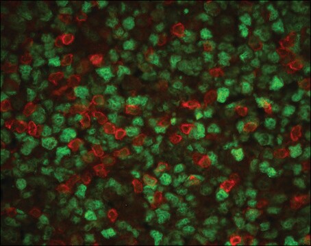
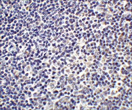
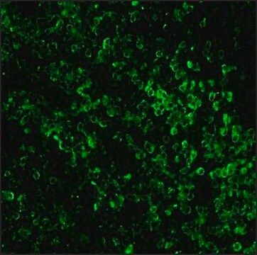
Anti-CD3ε (mouse) Antibody, PE, clone 145-2C11 is a highly specific armenian hamster monoclonal antibody, that targets CD3ε & has been tested in Flow Cytometry, IHC, Activity Assay, IP & western blotting.

Quality

The antibody was purified by affinity chromatography, and conjugated with PE under optimal conditions. The solution is free of unconjugated PE and unconjugated antibody.

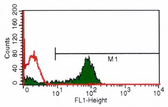
Flow Cytometry Analysis: 0.25 μg of this antibody detected CD3ε in one million C57BL/6 mouse splenocytes.

Target description

21 kDa calculated

Other Notes

Concentration: Please refer to the Certificate of Analysis for the lot-specific concentration.

Not finding the right product?  

Try our Product Selector Tool.

Storage Class Code

10 - Combustible liquids

WGK

WGK 2


Regulatory Listings

Regulatory Listings are mainly provided for chemical products. Only limited information can be provided here for non-chemical products. No entry means none of the components are listed. It is the user’s obligation to ensure the safe and legal use of the product.

JAN Code

MABF317:


Certificates of Analysis (COA)

Search for Certificates of Analysis (COA) by entering the products Lot/Batch Number. Lot and Batch Numbers can be found on a product’s label following the words ‘Lot’ or ‘Batch’.

Already Own This Product?

Find documentation for the products that you have recently purchased in the Document Library.

Visit the Document Library

M R Goodier et al.
Journal of immunology (Baltimore, Md. : 1950), 165(1), 139-147 (2000-06-22)
NK cells recognize and kill tumor cells and normal cells, and these play an important role in immune defense in cancer, infectious disease, and autoimmunity. NK killing is regulated by positive or negative signals derived from the interaction of surface
Wengang Wang et al.
Journal of immunology (Baltimore, Md. : 1950), 178(8), 4885-4890 (2007-04-04)
Sphingosine 1-phosphate (S1P) is a natural lipid mediator that regulates immune cell traffic, Ab production, and T cell cytokine generation by mechanisms that enhance Th2 activities. Responses to S1P are controlled principally by the diverse expression patterns of its receptors
Stephen L Tilley et al.
Journal of immunology (Baltimore, Md. : 1950), 178(5), 3208-3218 (2007-02-22)
The retinoid-related orphan receptors (ROR) comprise a distinct subfamily of nuclear receptors with the capacity to act as both repressors and activators of transcription. RORgamma, the most recently identified member of the ROR family, has been shown to be important
Fc receptor binding of anti-CD3 monoclonal antibodies is not essential for immunosuppression, but triggers cytokine-related side effects.
Vossen, A C, et al.
European Journal of Immunology, 25, 1492-1496 (1995)
Role of the programmed Death-1 pathway in the suppressive activity of alternatively activated macrophages in experimental cysticercosis.
Terrazas, Luis I, et al.
International Journal For Parasitology, 35, 1349-1358 (2005)

Our team of scientists has experience in all areas of research including Life Science, Material Science, Chemical Synthesis, Chromatography, Analytical and many others.

Contact Technical Service