Skip to Content
Merck
All Photos(4)

Key Documents

Safety Information

MABE327

Sigma-Aldrich

Anti-p53 (pantropic) Antibody, clone DO-1

clone DO-1, from mouse

Synonym(s):

Cellular tumor antigen p53, Antigen NY-CO-13, Phosphoprotein p53, Tumor suppressor p53

Sign Into View Organizational & Contract Pricing


About This Item

UNSPSC Code:
12352203
eCl@ss:
32160702
NACRES:
NA.41

biological source

mouse

Quality Level

antibody form

purified immunoglobulin

antibody product type

primary antibodies

clone

DO-1, monoclonal

species reactivity

human

technique(s)

immunocytochemistry: suitable
immunofluorescence: suitable
immunohistochemistry: suitable
immunoprecipitation (IP): suitable
western blot: suitable

isotype

IgG2aκ

NCBI accession no.

UniProt accession no.

shipped in

wet ice

target post-translational modification

unmodified

Gene Information

human ... TP53(7157)

General description

Cellular tumor antigen p53 (p53) is a ubiquitous transcription factor that is most widely known for its function as a tumor suppressor, through its regulation of cell growth and survival. p53 is activated in response to cell stress such as DNA damage and induces either cell cycle arrest or apoptosis. It coordinates the expression of multiple genes including p21, GADD45, Bax, and Puma. p53 is regulated by multiple mechanisms: At the post-transcriptional level, the stability of p53 mRNAs is regulated by the binding of molecules such as HuR, Wig-1, RPL26, L26, and WRAP53. Post-translationally, p53 undergoes phosphorylation and acetylation which may also play a role in regulating its activity. It may also undergo ubiquitination via the Akt-MDM2 pathway resulting in degradation by the 20S proteosome. Mutations in p53 are prevalent in many types of cancers, underscoring the therapeutic relevance of this protein.

Specificity

This antibody recognizes the N-terminus of p53 (pantropic).

Immunogen

Epitope: N-terminus
Recombinant protein corresponding to human p53 (pantropic).

Application

Immunocytochemistry Analysis: A 1:500 dilution from a representative lot detected p53 (pantropic) in A431 cells.

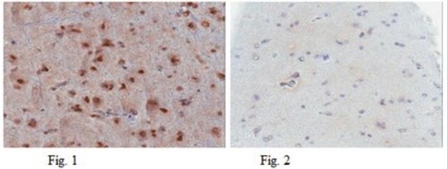
Immunohistochemistry Analysis: A 1:1,000 dilution from a representative lot detected p53 (pantropic) in human colorectal adenocarcinoma tissue and in human prostate cancer tissue.

Immunofluorescence Analysis: A 1:1,000 dilution from a representative lot detected p53 (pantropic) in human colorectal cancer cells.

Immunoprecipitation Analysis: A representative lot was used by an independent laboratory in radiolabelled HOS cell lysate (Vojtesek, B., et al. (1995). 71(6):1253-1256.).

Immunohistochemistry Analysis: A representative lot was used by an independent laboratory in primary breast carcinoma tissue (Beck, T., et al. (1995). Gynecol Oncol. 57(1):96-104.).
Research Category
Epigenetics & Nuclear Function
Research Sub Category
Cell Cycle, DNA Replication & Repair
Use Anti-p53 (pantropic) Antibody, clone DO-1 (Mouse Monoclonal Antibody) validated in WB, ICC, IHC, IF, IP to detect p53 (pantropic) also known as Cellular tumor antigen p53 or Antigen NY-CO-13.

Quality

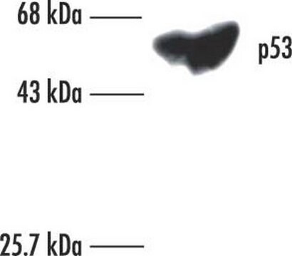
Evaluated by Western Blot in A431 cell lysate.

Western Blot Analysis: 1 µg/mL of this antibody detected p53 (pantropic) in 10 µg of A431 cell lysate. This antibody detects isoforms 1, 2, and 3.

Target description

~53 kDa observed. Uniprot describes 9 different isoforms due to alternative splicing and post-translational modification. This antibody detects isoform 1 (~38 kDa), isoform 2 (~39 kDa), and isoform 3 (~39 kDa). An uncharacterized band may be observed at ~43 kDa in some cell lysates.

Linkage

Replaces: 04-1083

Physical form

Format: Purified
Protein G Purified
Purified mouse monoclonal IgG2aκ in buffer containing 0.1 M Tris-Glycine (pH 7.4), 150 mM NaCl with 0.05% sodium azide.

Storage and Stability

Stable for 1 year at 2-8°C from date of receipt.

Analysis Note

Control
A431 cell lysate

Other Notes

Concentration: Please refer to the Certificate of Analysis for the lot-specific concentration.

Disclaimer

Unless otherwise stated in our catalog or other company documentation accompanying the product(s), our products are intended for research use only and are not to be used for any other purpose, which includes but is not limited to, unauthorized commercial uses, in vitro diagnostic uses, ex vivo or in vivo therapeutic uses or any type of consumption or application to humans or animals.

Not finding the right product?  

Try our Product Selector Tool.

Storage Class Code

12 - Non Combustible Liquids

WGK

WGK 1

Flash Point(F)

Not applicable

Flash Point(C)

Not applicable


Regulatory Listings

Regulatory Listings are mainly provided for chemical products. Only limited information can be provided here for non-chemical products. No entry means none of the components are listed. It is the user’s obligation to ensure the safe and legal use of the product.

JAN Code

MABE327:


Certificates of Analysis (COA)

Search for Certificates of Analysis (COA) by entering the products Lot/Batch Number. Lot and Batch Numbers can be found on a product’s label following the words ‘Lot’ or ‘Batch’.

Already Own This Product?

Find documentation for the products that you have recently purchased in the Document Library.

Visit the Document Library

B Vojtesek et al.
British journal of cancer, 71(6), 1253-1256 (1995-06-01)
We analysed antibodies specific for human p53 in sera from primary breast cancer patients using three different immunoassays and we related these results to the p53 level in tumour tissue detected by immunohistochemistry. Only 44% (11/25) of apparently enzyme-linked immunosorbent
Kerstin Herzer et al.
Cancer research, 65(23), 10830-10837 (2005-12-03)
Tumor suppressor protein promyelocytic leukemia (PML) is implicated in apoptosis regulation and antiviral response. PML localizes predominantly to PML-nuclear bodies (PML-NB), nuclear macromolecular complexes regulating tumor suppressor protein p53 activity. Consistent with the function of PML in the cellular antiviral
Bo Yun Lee et al.
Aging cell, 5(2), 187-195 (2006-04-22)
Replicative senescence limits the proliferation of somatic cells passaged in culture and may reflect cellular aging in vivo. The most widely used biomarker for senescent and aging cells is senescence-associated beta-galactosidase (SA-beta-gal), which is defined as beta-galactosidase activity detectable at
Usefulness of immunohistochemical staining for p53 in the prognosis of breast carcinomas: correlations with established prognosis parameters and with the proliferation marker, MIB-1.
Beck, T, et al.
Gynecologic Oncology, 57, 96-104 (1995)
June Li et al.
Carcinogenesis, 28(6), 1163-1170 (2007-02-06)
Cells from patients with genomic instability syndromes have high predisposition to cancer. However, little is known about whether these mutant cells have high susceptibility to oncogenic transformation. We have tested the hypothesis that a defect in maintaining genome integrity is

Our team of scientists has experience in all areas of research including Life Science, Material Science, Chemical Synthesis, Chromatography, Analytical and many others.

Contact Technical Service