Skip to Content
Merck
All Photos(4)

Key Documents

Safety Information

MABC46

Sigma-Aldrich

Anti-Atg13 Antibody, clone 2H4.2

clone 2H4.2, from mouse

Synonym(s):

Autophagy-related protein 13

Sign Into View Organizational & Contract Pricing


About This Item

UNSPSC Code:
12352203
eCl@ss:
32160702
NACRES:
NA.41
clone:
2H4.2, monoclonal
application:
IHC
WB
species reactivity:
human
technique(s):
immunohistochemistry: suitable
western blot: suitable
citations:
8

biological source

mouse

Quality Level

antibody form

purified immunoglobulin

antibody product type

primary antibodies

clone

2H4.2, monoclonal

species reactivity

human

technique(s)

immunohistochemistry: suitable
western blot: suitable

isotype

IgG1κ

NCBI accession no.

UniProt accession no.

shipped in

wet ice

target post-translational modification

unmodified

Gene Information

human ... ATG13(9776)

General description

The autophagy-related protein 13 (Atg13) plays an important role in the formation of autophagosomes. Autophagosomes are formed in response to nutrient deprivation and function as the transport vesicles for organelles, proteins, and protein complexes targeted for lysosomes that digest these cargos to produce energy and nutrients. Atg13 is activated by the mTOR pathway and forms a complex with the FIP200 protein. This complex is involved in enhancing the activity of the ULK1 kinase which is required for the formation of autophagosomes. Atg13/FIP200 faciliates the localization of ULK1 to pre-autophagosomes, and subsequently stabilizes ULK1. Autophagy is an important process in development, growth, and cell differentiation, and disruption of this process may contribute to cancer, aging, and neurodegenerative diseases.

Immunogen

GST-tagged recombinant protein corresponding to human Atg13.

Application

Research Category
Apoptosis & Cancer
Research Sub Category
Apoptosis - Additional
This Anti-Atg13 Antibody, clone 2H4.2 is validated for use in Western Blotting, IHC for the detection of Atg13.
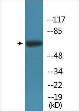
Western Blot Analysis: A representative lot detected Atg13 in HeLa cell lysate.

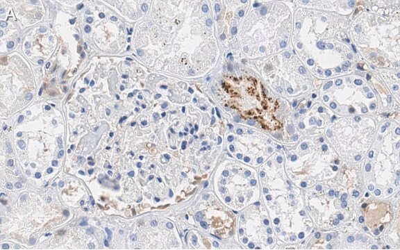
Immunohistochemistry Analysis: A 1:50 dilution from a representative lot detected Atg13 in normal human substancia nigra tissues.

Quality

Evaluated by Western Blot in campthothecin treated HeLa cell lysate.

Western Blot Analysis: A 1:1,000 dilution of this antibody detected Atg13 in 10 µg of campthothecin treated HeLa cell lysate.

Target description

~60 kDa observed

Physical form

Format: Purified
Protein G Purified
Purified mouse monoclonal IgG1κ in buffer containing 0.1 M Tris-Glycine (pH 7.4), 150 mM NaCl with 0.05% sodium azide.

Storage and Stability

Stable for 1 year at 2-8°C from date of receipt.

Analysis Note

Control
Campthothecin treated HeLa cell lysate.

Disclaimer

Unless otherwise stated in our catalog or other company documentation accompanying the product(s), our products are intended for research use only and are not to be used for any other purpose, which includes but is not limited to, unauthorized commercial uses, in vitro diagnostic uses, ex vivo or in vivo therapeutic uses or any type of consumption or application to humans or animals.

Not finding the right product?  

Try our Product Selector Tool.

Storage Class Code

12 - Non Combustible Liquids

WGK

WGK 1

Flash Point(F)

Not applicable

Flash Point(C)

Not applicable


Regulatory Listings

Regulatory Listings are mainly provided for chemical products. Only limited information can be provided here for non-chemical products. No entry means none of the components are listed. It is the user’s obligation to ensure the safe and legal use of the product.

JAN Code

MABC46:


Certificates of Analysis (COA)

Search for Certificates of Analysis (COA) by entering the products Lot/Batch Number. Lot and Batch Numbers can be found on a product’s label following the words ‘Lot’ or ‘Batch’.

Already Own This Product?

Find documentation for the products that you have recently purchased in the Document Library.

Visit the Document Library

Yo-Hei Yamamoto et al.
The Journal of cell biology, 219(8) (2020-06-04)
In macroautophagy, membrane structures called autophagosomes engulf substrates and deliver them for lysosomal degradation. Autophagosomes enwrap a variety of targets with diverse sizes, from portions of cytosol to larger organelles. However, the mechanism by which autophagosome size is controlled remains
Marianna Carinci et al.
The EMBO journal, 40(10), e103563-e103563 (2021-05-02)
The early secretory pathway and autophagy are two essential and evolutionarily conserved endomembrane processes that are finely interlinked. Although growing evidence suggests that intracellular trafficking is important for autophagosome biogenesis, the molecular regulatory network involved is still not fully defined.
Suresh Kumar et al.
Developmental cell, 49(1), 130-144 (2019-03-05)
Syntaxin 17 (Stx17) has been implicated in autophagosome-lysosome fusion. Here, we report that Stx17 functions in assembly of protein complexes during autophagy initiation. Stx17 is phosphorylated by TBK1 whereby phospho-Stx17 controls the formation of the ATG13+FIP200+ mammalian pre-autophagosomal structure (mPAS)
Cindy Puente et al.
The Journal of biological chemistry, 291(11), 6026-6035 (2016-01-24)
Autophagy is a conserved catabolic process that utilizes a defined series of membrane trafficking events to generate a de novo double-membrane vesicle termed the autophagosome, which matures by fusing to the lysosome. Subsequently, the lysosome facilitates the degradation and recycling

Our team of scientists has experience in all areas of research including Life Science, Material Science, Chemical Synthesis, Chromatography, Analytical and many others.

Contact Technical Service