Skip to Content
Merck
All Photos(1)

Key Documents

MAB5266

Sigma-Aldrich

Anti-Neurofilament 200 kDa Antibody, clone N52

clone N52, Chemicon®, from mouse

Synonym(s):

Anti-CMT2CC, Anti-NFH

Sign Into View Organizational & Contract Pricing


About This Item

UNSPSC Code:
12352203
eCl@ss:
32160702
NACRES:
NA.41

biological source

mouse

Quality Level

100
300

antibody form

purified immunoglobulin

antibody product type

primary antibodies

clone

N52, monoclonal

species reactivity

human, mouse, monkey, rat, pig

manufacturer/tradename

Chemicon®

technique(s)

immunohistochemistry: suitable
western blot: suitable

isotype

IgG1

NCBI accession no.

UniProt accession no.

shipped in

wet ice

target post-translational modification

unmodified

Gene Information

human ... NEFH(4744)
mouse ... Nefh(380684)
pig ... Nefh(100156492)
rat ... Nefh(24587)
rhesus monkey ... Nefh(717705)

General description

Neurofilaments are a type of intermediate filament that serve as major elements of the cytoskeleton supporting the axon cytoplasm. They are the most abundant fibrillar components of the axon, being on average 3-10 times more frequent than axonal microtubules. Neurofilaments (10nm in dia.) are built from three intertwined protofibrils which are themselves composed of two tetrameric protofilament complexs of monomeric proteins. The neurofilament triplet proteins (68/70, 160, and 200 kDa) occur in both the central and peripheral nervous system and are usually neuron specific. The 68/70 kDa NF-L protein can self-assemble into a filamentous structure, however the 160 kDa NF-M and 200 kDa NF-H proteins require the presence of the 68/70 kDa NF-L protein to co-assemble. Neuromas, ganglioneuromas, gangliogliomas, ganglioneuroblastomas and neuroblastomas stain positively for neurofilaments. Although typically restricted to neurons, neurofilaments have been detected in paragangliomas and adrenal and extra-adrenal pheochromocytomas. Carcinoids, neuroendocrine carcinomas of the skin, and oat cell carcinomas of the lung also express neurofilaments. For more neurofilament information see Nervous System Cell Type Specific Marker chart online under the CHEMICON Technical Support section.

Specificity

MAB5266 reacts with the phophorylated and dephosphorylated H-chain of neurofilament 200kDa (NF-H) in normal tissues/extracts (Shaw, 1986). MAB5266 can be used to detect cells of neuronal origin by immunohistochemistry and Western blot. It has been reported that reactivity of MAB5266 with NF-H is blocked in cdk-5 over-expressing cells (Guidato, 1996).

Immunogen

Carboxy terminal tail segment of enzymatically dephosphorylated porcine H-chain.

Application

Research Category
Neuroscience
Research Sub Category
Neurofilament & Neuron Metabolism

Neuronal & Glial Markers
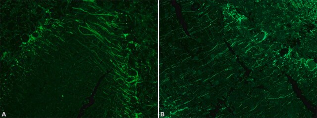
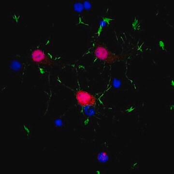
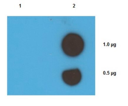
This Anti-Neurofilament 200 kDa Antibody, clone N52 is validated for use in WB, IH for the detection of Neurofilament 200 kDa.
Western blot: 5-10 μg/mL

Immunohistochemistry: 5-10 μg/mL

Optimal working dilutions must be determined by end user.

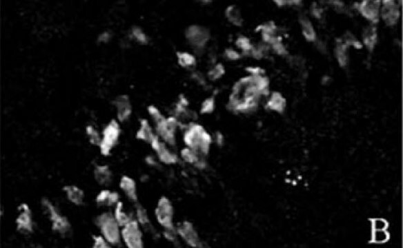
Immunohistochemistry: Antibody N52 reacts with a fragment of NF-200 side-arm that is located at the end of the MPR KSP domain and which contains the consensus cdk-5 phosphorylation site, however this reactivity is abolished if the NF-200 fragment becomes phosphorylated by cdk-5 {Guidato et al, 1996}. Thus for the fullest staining and reactivity, including westerns, it is suggested that samples be treated with alkaline phosphatase prior to antibody staining. The following treatment protocol is suggested:


ProtocolPreparation of Solutions:

I. TBS: Tris-HCl, 0.1 mol/l; NaCl, 0.1 mol/l′ pH 8.0.

II. Mix alkaline phosphatase, 1 mg/ml with 1 mol/l ZnSO4,; 1mol/l MgCl2, and 1mmol/l PMSF; and dissove in TBS.

NOTE: It is necessary to dialyze the alkaline phosphatase against solution I before use.

Procedure:

Frozen sections from shock frozen tissue samples are air-dried and then fixed with acetone for 10 min at -20°C. The excess acetone is allowed to evaporate at room temperature. Incubate the sections in solutions II for 4 hours at +30oC and wash in TBS 3 times for 5 minutes each. Negative control sections are incubated in solution II with out alkaline phosphatase. Further treatment as follows:

- Cover the preparation with 10-20 μl antibody solution and incubate for 1 hr in a humid chamber.

- Immerse the slide in TBS and wash in TBS 3 times for 5 minutes each.

- Cover the preparation with 10-20 μl of a solution of anti-mouse Ig-antibody, labeled with fluorscine isothiocyanate, and incubate in a humid chamber at 37°C for 1 hour.

- Wash the slide in TBS 3 times for 5 min each.

Physical form

Format: Purified
Purified immunoglobulin (Ion exchange chromatography). Liquid in 0.02M phosphate buffer, 0.25M NaCl with 0.1% sodium azide, pH 7.6

Storage and Stability

Maintain at 2-8°C in undiluted aliquots for up to 6 months. Avoid repeated freeze/thaw cycles.

Other Notes

Concentration: Please refer to the Certificate of Analysis for the lot-specific concentration.

Legal Information

CHEMICON is a registered trademark of Merck KGaA, Darmstadt, Germany

Disclaimer

Unless otherwise stated in our catalog or other company documentation accompanying the product(s), our products are intended for research use only and are not to be used for any other purpose, which includes but is not limited to, unauthorized commercial uses, in vitro diagnostic uses, ex vivo or in vivo therapeutic uses or any type of consumption or application to humans or animals.

Not finding the right product?  

Try our Product Selector Tool.

Storage Class Code

10 - Combustible liquids

WGK

WGK 2

Flash Point(F)

Not applicable

Flash Point(C)

Not applicable


Certificates of Analysis (COA)

Search for Certificates of Analysis (COA) by entering the products Lot/Batch Number. Lot and Batch Numbers can be found on a product’s label following the words ‘Lot’ or ‘Batch’.

Already Own This Product?

Find documentation for the products that you have recently purchased in the Document Library.

Visit the Document Library

Customers Also Viewed

Ami V Patel et al.
Developmental biology, 340(2), 419-429 (2010-02-04)
In mice lacking functional brain-derived neurotrophic factor (BDNF), the number of geniculate ganglion neurons, which innervate taste buds, is reduced by one-half. Here, we determined how and when BDNF regulates the number of neurons in the developing geniculate ganglion. The
Phosphorylation of neurofilament H subunit as related to arrangement of neurofilaments.
Gotow, T and Tanaka, J
Journal of Neuroscience Research, 37, 691-713 (1994)
Sheona Watson-Scales et al.
PLoS genetics, 14(5), e1007383-e1007383 (2018-05-11)
Down Syndrome (DS) is caused by trisomy of chromosome 21 (Hsa21) and results in a spectrum of phenotypes including learning and memory deficits, and motor dysfunction. It has been hypothesized that an additional copy of a few Hsa21 dosage-sensitive genes
Jake A Robinson et al.
The American journal of pathology, 190(7), 1530-1544 (2020-04-05)
HIV-associated sensory neuropathy is a common neurologic comorbidity of HIV infection and prevails in the post-antiretroviral therapy (ART) era. HIV infection drives pathologic changes in the dorsal root ganglia (DRG) through inflammation, altered metabolism, and neuronal dysfunction. Herein, we characterized
Effects of spinal nerve ligation on immunohistochemically identified neurons in the L4 and L5 dorsal root ganglia of the rat.
Hammond, Donna L, et al.
The Journal of Comparative Neurology, 475, 575-589 (2004)

Our team of scientists has experience in all areas of research including Life Science, Material Science, Chemical Synthesis, Chromatography, Analytical and many others.

Contact Technical Service