Skip to Content
Merck
All Photos(2)

Key Documents

Safety Information

MAB5258-I

Sigma-Aldrich

Anti-Synaptophysin Antibody, clone SY38

clone SY38, from mouse

Synonym(s):

Synaptophysin, Major synaptic vesicle protein p38

Sign Into View Organizational & Contract Pricing


About This Item

UNSPSC Code:
12352203
eCl@ss:
32160702
NACRES:
NA.41

biological source

mouse

Quality Level

antibody form

purified immunoglobulin

antibody product type

primary antibodies

clone

SY38, monoclonal

species reactivity

bovine, mouse, rat, human

technique(s)

immunocytochemistry: suitable
immunofluorescence: suitable
immunohistochemistry: suitable (paraffin)
western blot: suitable

isotype

IgG1κ

NCBI accession no.

UniProt accession no.

shipped in

ambient

target post-translational modification

unmodified

Gene Information

human ... SYP(6855)

General description

Synaptophysin (UniProt P20488; also known as Major synaptic vesicle protein p38) is encoded by the SYP gene (Gene ID 280937) in bovine species. Synaptophysin is a 38 kDa four-transmembrane glycoprotein on synaptic vesicle (SV) with both its N- and C-termini exposed cytoplasmically. Synaptophysin forms homomultimers in the SV membrane and is heavily phosphorylated on tyrosine residues in its C-terminal pentapeptide repeats. Synapthophysin is also known to interact with SV v-SNARE protein synaptobrevin II (sybII) when sybII is not engaged with other SNARE proteins, suggesting a role of synaptophysin in sybII retrieval during SV endocytosis. Consistently, synaptophysin-knockout neurons display reduced level of sybII at their nerve terminals.

Specificity

Clone SY38 detects synaptophysin and stains synaptophysin-containing vesicles by targeting the flexible segment -SGGGG- in the center of the C-termimal pentapeptide repeats (Knaus, P., and Betz, H. (1990). FEBS Lett. 261(2):358-360).

Immunogen

Presynaptic vesicles from bovine brain (Wiedenmann, B., and Franke, W.W. (1985). Cell. 41(3):1017-1028).

Application

Anti-Synaptophysin, clone SY38, Cat. No. MAB5258-I, is a highly specific mouse monoclonal antibody that targets Synaptophysin and has been tested in Immunocytochemistry, Immunofluorescence, Immunohistochemistry, and Western Blotting.
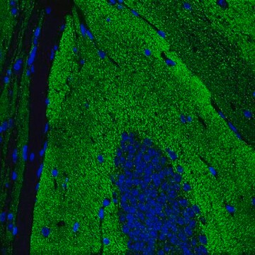
Immunohistochemistry Analysis: A 1:50 dilution from a representative lot detected Synaptophysin in human cerebellum and cerebral cortex tissue sections.

Western Blotting Analysis: A 1:1,000 dilution from a representative lot detected Synaptophysin in 10 µg of rat hippocampus and human whole brain tissue lysates.

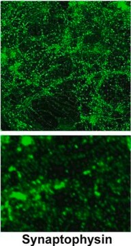
Immunocytochemistry Analysis: Representative lots immunostained presynaptic vesicles of 4% formaldehyde-fxied, 0.2% Triton X-100-permeabilzied primary mouse hippocampal neurons by fluorescent immunocytochemistry (Hu, X., et al. (2008). J. Neurosci. 28(49):13094-13105; Tarr, P.T., and Edwards, P.A. (2008). J. Lipid Res. 49(1):169-182).

Immunofluorescence Analysis: A representative lot immunostained presynaptic membrane of neurons by fluorescent immunohistochemistry staining of 4% paraformaldehyde-fixed, 0.3% Triton X-100-permeabilized, OCT-embedded rat spinal cord cryosections (Stück, E.D., et al. (2012). Neural Plast. 2012:261345).

Immunofluorescence Analysis: A representative lot immunostained synaptophysin around a large neuron in the ventral horn of the lumbar (L3/L4) segment by fluorescent immunohistochemistry staining of 2% paraformaldehyde/0.2% parabenzoquinone-fixed free-floating rat spinal cord sections (Macias, M., et al. (2009). BMC Neurosci. 10:144).

Immunofluorescence Analysis: A representative lot immunostained synaptophysin in bovine (pancreas) and human (pancreas, pheochromocytoma, and islet-cell carcinoma) frozen tissue sections (Wiedenmann, B., et al. (1986). Proc. Natl. Acad. Sci. U.S.A. 83(10):3500-3504).

Western Blotting Analysis: A representative lot detected an upregulated synaptophysin expression in human iPSCs with a 4-bp deletion in DISC1 gene (Wen, Z., et al. (2014). Nature. 515(7527):414-418).

Western Blotting Analysis: A representative lot detected synaptophysin distribution among PC12 rat pheochromocytoma cell membrane fractions (Salazar, G., et al. (2005). Mol. Biol. Cell. 16(8):3692-3704).

Western Blotting Analysis: A representative lot detected only synaptophysin recombinant contructs that contained the flexible segment -SGGGG- in the center of the c-terminal pentapeptide repeats (Knaus, P., and Betz, H. (1990). FEBS Lett. 261(2):358-360).
Research Category
Neuroscience

Quality

Evaluated by Western Blotting in mouse hypothalamus tissue lysate.

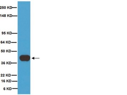
Western Blotting Analysis: A 1:1,000 dilution of this antibody detected Synaptophysin in 10 µg of mouse hypothalamus tissue lysate.

Target description

~38 kDa observed. 33.91/34.03/33.31 kDa (bovine/mouse/rat), 33.85/20.76 kDa (human isoform 1/2) calculated. Uncharacterized bands may be observed in some lysate(s).

Physical form

Format: Purified
Protein G purified.
Purified mouse IgG1κ in buffer containing 0.1 M Tris-Glycine (pH 7.4), 150 mM NaCl with 0.05% sodium azide.

Storage and Stability

Stable for 1 year at 2-8°C from date of receipt.

Other Notes

Concentration: Please refer to lot specific datasheet.

Disclaimer

Unless otherwise stated in our catalog or other company documentation accompanying the product(s), our products are intended for research use only and are not to be used for any other purpose, which includes but is not limited to, unauthorized commercial uses, in vitro diagnostic uses, ex vivo or in vivo therapeutic uses or any type of consumption or application to humans or animals.

Not finding the right product?  

Try our Product Selector Tool.

Storage Class Code

12 - Non Combustible Liquids

WGK

WGK 1


Regulatory Listings

Regulatory Listings are mainly provided for chemical products. Only limited information can be provided here for non-chemical products. No entry means none of the components are listed. It is the user’s obligation to ensure the safe and legal use of the product.

JAN Code

MAB5258-I:


Certificates of Analysis (COA)

Search for Certificates of Analysis (COA) by entering the products Lot/Batch Number. Lot and Batch Numbers can be found on a product’s label following the words ‘Lot’ or ‘Batch’.

Already Own This Product?

Find documentation for the products that you have recently purchased in the Document Library.

Visit the Document Library

Jennetta W Hammond et al.
Glia, 66(12), 2563-2574 (2018-10-17)
Microglial activation, increased proinflammatory cytokine production, and a reduction in synaptic density are key pathological features associated with HIV-associated neurocognitive disorders (HAND). Even with combination antiretroviral therapy (cART), more than 50% of HIV-positive individuals experience some type of cognitive impairment.
Jiao-Jiao Yang et al.
Molecular and clinical oncology, 12(2), 148-154 (2020-01-14)
A 55-year-old woman presented with chest and back pain of unknown cause. Contrast-enhanced computed tomography revealed two low-density tumors, sized 4.6 and 4.4 cm, in the hepatic caudate and left inner lobes, respectively. There are multiple enlarged lymph nodes around
Qifa Li et al.
Frontiers in molecular neuroscience, 12, 26-26 (2019-03-12)
Chondroitin sulfate (CS) and dermatan sulfate (DS) proteoglycans (PGs) are major extracellular matrix (ECM) components of the central nervous system (CNS). A large body of evidence has shown that CSPGs/DSPGs play critical roles in neuronal growth, axon guidance, and plasticity
Paul Lu et al.
Stem cell reports, 13(1), 105-114 (2019-06-18)
Neural progenitor cells (NPCs) transplanted into sites of spinal cord injury (SCI) extend large numbers of axons into the caudal host spinal cord. We determined the precise locations of neurons in the graft that extend axons into the caudal host
Tomomi Kiyota et al.
Journal of neuroimmunology, 319, 80-92 (2018-03-27)
We investigated the effects of granulocyte-macrophage colony stimulating factor (GM-CSF) on behavioral and pathological outcomes in Alzheimer's disease (AD) and non-transgenic mice. GM-CSF treatment in AD mice reduced brain amyloidosis, increased plasma Aβ, and rescued cognitive impairment with increased hippocampal

Our team of scientists has experience in all areas of research including Life Science, Material Science, Chemical Synthesis, Chromatography, Analytical and many others.

Contact Technical Service