MAB3568
Anti-Myosin Antibody, smooth muscle heavy chain, SM1 & SM2, clone ID8
clone ID8 (SMMS-1), Chemicon®, from mouse
Synonym(s):
Anti-CCDC128, Anti-KLRAQ1
About This Item
Recommended Products
biological source
mouse
Quality Level
antibody form
purified immunoglobulin
antibody product type
primary antibodies
clone
ID8 (SMMS-1), monoclonal
species reactivity
human, bovine, pig, rabbit
manufacturer/tradename
Chemicon®
technique(s)
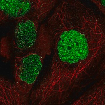
immunocytochemistry: suitable
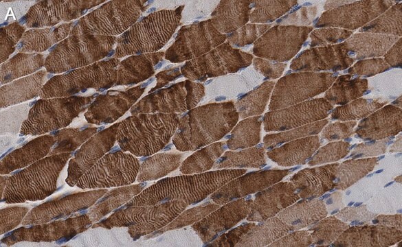
immunohistochemistry: suitable (paraffin)
immunoprecipitation (IP): suitable
western blot: suitable
isotype
IgG1
NCBI accession no.
UniProt accession no.
shipped in
wet ice
target post-translational modification
unmodified
Gene Information
human ... MYH11(4629)
Application
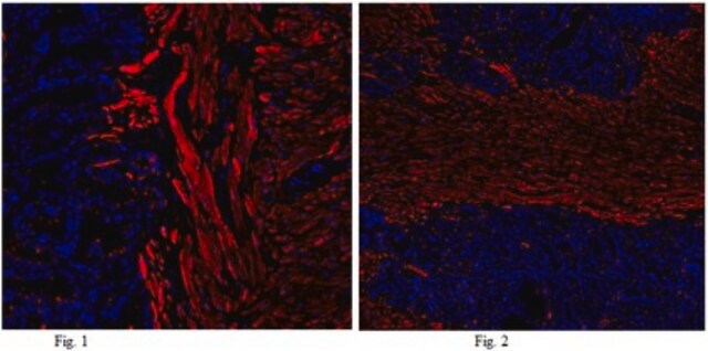
Immunohistochemistry on frozen and paraffin embedded tissue sections. Suggested fixation for frozen tissue sections is acetone fix for 6 minutes at room temperature. For formalin fixed paraffin embedded tissue sections: microwave in 0.01M citrate buffer (pH 6.0) for 8-10 minutes (note that all microwaves differ and adjustments may need to be made) follow with enzyme digestion (0.01% pronase for 10 mintues). Suggested blocking agent is fetal bovine serum.
Immunocytochemistry
Immunoprecipitation. Suggested extraction buffer is 20 mM Tris-HCl, pH 7.4, 150 mM NaCl, 1% Triton X-100, 0.1% SDS, 0.5% deoxycholic acid-NaCl and 0.5 mM PMSF. Final reaction volume is 1 mL and suggested capture agent is agarose conjugated anti-mouse IgG.
Optimal working dilutions must be determined by the end user.
APPLICATION NOTES FOR MAB3568
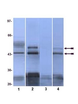
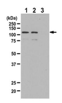
WESTERN BLOT
To achieve good resolution of myosin heavy chain isoforms with distinct molecular weight (200 - 2004 kDa), the following procedure should be followed: 1). Pyrophoshate extraction buffer for sample preparation (see below); run SDS-PAGE in 5% gel. Important: for better resolution of the MHC bands, use electrophoretic buffer with pH 8.2 (i.e. 0.1 less than standard), and prepare resolving gel (5%) with pH 9.0 (not 8.8 as usual). Also help thorough degasing of the resolving gel mixture (H2O, acrylamide, EDTA, pH 9.0, before (!) adding SDS, TEMED and APS). Run SDS-PAGE longer than after the dye front runs off (use 200 kDa MW markers and let it′s 200 kDa band run at least to the middle of 8X8 gel (using a big size gel (not the mini-gel!) will enhance the quality of MHC band resolution).
Pyrophosphate extraction buffer: (40mM Na4P2O7x10H2O, 1mM MgCl2, 1mM EGTA (add KOH to dissolve EGTA), PMSF, pH 9.5). To extract acto-myosin from tissues/cells, shake minced tissue or cells in cold extraction buffer 1 hr on ice bath (0oC), centrifuge @10,000g for 10 min at +2-8 oC, take supernatant and mix it 1:1 with standard Laemmli sample buffer, boil, run SDS-PAGE in 5% gel (see above).
Physical form
Analysis Note
POSITIVE CONTROL: Smooth muscle (e.g. aterial tunica media). Negative control: any nonmuscle tissue (e.g. arterial tunica adentitial).
Other Notes
Legal Information
Not finding the right product?
Try our Product Selector Tool.
Storage Class Code
10 - Combustible liquids
WGK
WGK 2
Flash Point(F)
Not applicable
Flash Point(C)
Not applicable
Regulatory Listings
Regulatory Listings are mainly provided for chemical products. Only limited information can be provided here for non-chemical products. No entry means none of the components are listed. It is the user’s obligation to ensure the safe and legal use of the product.
JAN Code
MAB3568:
Certificates of Analysis (COA)
Search for Certificates of Analysis (COA) by entering the products Lot/Batch Number. Lot and Batch Numbers can be found on a product’s label following the words ‘Lot’ or ‘Batch’.
Already Own This Product?
Find documentation for the products that you have recently purchased in the Document Library.
Our team of scientists has experience in all areas of research including Life Science, Material Science, Chemical Synthesis, Chromatography, Analytical and many others.
Contact Technical Service