Skip to Content
Merck
All Photos(3)

Key Documents

Safety Information

MAB1273B

Sigma-Aldrich

Anti-Mitochondria Antibody, clone 113-1, Biotin Conjugate

clone 113-1, from mouse, biotin conjugate

Synonym(s):

Human Mitochondria

Sign Into View Organizational & Contract Pricing


About This Item

UNSPSC Code:
12352203
eCl@ss:
32160702
NACRES:
NA.41

biological source

mouse

Quality Level

conjugate

biotin conjugate

antibody form

purified immunoglobulin

antibody product type

primary antibodies

clone

113-1, monoclonal

species reactivity

human (mitochondria protein)

should not react with

rat (mitochondria protein), mouse (mitochondria protein)

technique(s)

immunocytochemistry: suitable
immunohistochemistry: suitable

isotype

IgG1

shipped in

wet ice

target post-translational modification

unmodified

General description

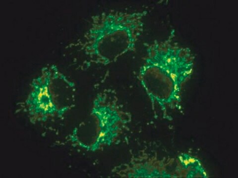
This antibody recognizes a 60 kDa nonglycosylated protein component of mitochondria found in human cells. It can be used to stain mitochondria in cell, tissue or biochemical preparations and can be used as a marker of the mitochondria in subcellular fractions. MAB1273B recognizes a 65 kD protein by immunoprecipitation and produces the stringy, "spaghetti-like", staining patten in the cytoplasm of human cells.

Specificity

This antibody recognizes human. Antibody is not reactive with mouse or rodent.

Immunogen

Surface of intact human mitochondria

Application

Detect Mitochondrial Marker using this mouse monoclonal antibody, Anti-Mitochondria Antibody, clone 113-1, Biotin Conjugate validated for use in ICC & IHC.
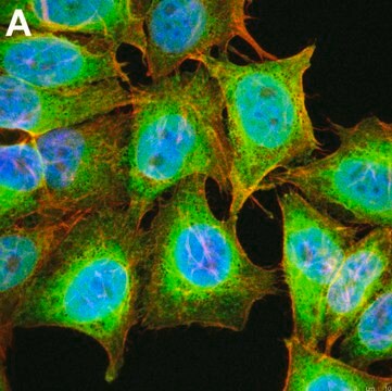
Evaluated by Immunocytochemistry in primary mouse embryonic fibroblasts (PMEFs).
Immunocytochemsitry Analysis: A 1:50 dilution of this antibody did not detect mitrochondria in primary mouse embryonic fibroblasts (PMEFs). Evaluated by Immunohistochemistry in human kidney tissue.
Immunocytochemsitry Analysis: A 1:500 dilution of this antibody detected mitrochondria in human kidney tissue.
Research Category
Stem Cell Research

Cell Structure
Research Sub Category
Developmental Neuroscience

Organelle & Cell Markers

Quality

Evaluated by Immunocytochemistry in human adipose mesenchymal stem cells (SCC038).
Immunocytochemsitry Analysis: A 1:50 dilution of this antibody detected mitrochondria in human adipose mesenchymal stem cells (SCC038).

Target description

60 kDa calculated

Physical form

Protein G Purified
Purified mouse monoclonal IgG1 conjugated to Biotin in PBS with 0.1% sodium azide and 15mg/ml BSA.

Storage and Stability

Maintain refrigerated at 2-8ºC protected from light in undiluted aliquots for up to 6 months from date of receipt.

Analysis Note

Control
Human adipose mesenchymal stem cells (SCC038).

Other Notes

Concentration: Please refer to the Certificate of Analysis for the lot-specific concentration.

Disclaimer

Unless otherwise stated in our catalog or other company documentation accompanying the product(s), our products are intended for research use only and are not to be used for any other purpose, which includes but is not limited to, unauthorized commercial uses, in vitro diagnostic uses, ex vivo or in vivo therapeutic uses or any type of consumption or application to humans or animals.

Not finding the right product?  

Try our Product Selector Tool.

Storage Class Code

12 - Non Combustible Liquids

WGK

WGK 2

Flash Point(F)

Not applicable

Flash Point(C)

Not applicable


Regulatory Listings

Regulatory Listings are mainly provided for chemical products. Only limited information can be provided here for non-chemical products. No entry means none of the components are listed. It is the user’s obligation to ensure the safe and legal use of the product.

JAN Code

MAB1273B:


Certificates of Analysis (COA)

Search for Certificates of Analysis (COA) by entering the products Lot/Batch Number. Lot and Batch Numbers can be found on a product’s label following the words ‘Lot’ or ‘Batch’.

Already Own This Product?

Find documentation for the products that you have recently purchased in the Document Library.

Visit the Document Library

Emily Pulford et al.
Pathology, 48(7), 650-659 (2016-12-14)
Vasculogenic mimicry, the process in which cancer cells form angiomatoid structures independent of or in addition to host angiogenesis has been recorded in several otherwise non-endothelial malignant neoplasms. This study describes evidence of routine vascular mimicry by human mesothelioma cell
Kai Kang et al.
Biomaterials, 230, 119574-119574 (2019-11-26)
Engineered heart tissues (EHTs) are regarded as being the most promising alternative to synthetic materials, and autologous mesenchymal stem cells (MSCs) are widely used as seeding cells. However, few studies have evaluated the feasibility of using MSCs from patients with

Our team of scientists has experience in all areas of research including Life Science, Material Science, Chemical Synthesis, Chromatography, Analytical and many others.

Contact Technical Service