Skip to Content
Merck
All Photos(2)

Key Documents

Safety Information

ABT29

Sigma-Aldrich

Anti-Tensin-3 Antibody

from rabbit, purified by affinity chromatography

Synonym(s):

Tensin-3, Tensin-like SH2 domain-containing protein 1, Tumor endothelial marker 6

Sign Into View Organizational & Contract Pricing


About This Item

UNSPSC Code:
12352203
eCl@ss:
32160702
NACRES:
NA.41

biological source

rabbit

Quality Level

antibody form

affinity isolated antibody

antibody product type

primary antibodies

clone

polyclonal

purified by

affinity chromatography

species reactivity

human

technique(s)

immunocytochemistry: suitable
western blot: suitable

NCBI accession no.

UniProt accession no.

shipped in

wet ice

target post-translational modification

unmodified

Gene Information

human ... TNS3(64759)

General description

Tensin-3 is a member of the Tensin family of proteins that serve as a connection between the cytoskeleton and extracellular matrix. Because of this link, Tensin’s are commonly thought to regulate signaling for the shape of the cell and motility. Tensin-3 localizes in the cell cytoplasm and has been shown to be significantly decreased in many different human cancer cell lines. Higher expression levels of Tensin-3 were also found to be associated with longer survival. Tensin-3 has also been found to be highly expressed in functioning thyroid follicular adenomas while expression is significantly lower in non-functioning thyroid follicular adenomas, which seems to imply that Tensin-3 has an important role in basic thyroid function.

Immunogen

KLH-conjugated linear peptide corresponding to human Tensin-3.

Application

Anti-Tensin-3 Antibody detects level of Tensin-3 & has been published & validated for use in WB & IC.
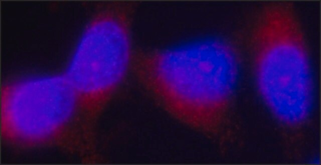
Immunocytochemistry Analysis: A 1:500 dilution from a representative lot detected Tensin-3 in A431 cells.
Research Category
Cell Structure
Research Sub Category
ECM Proteins

Cytoskeleton

Quality

Evaluated by Western Blot in human kidney tissue lysate.

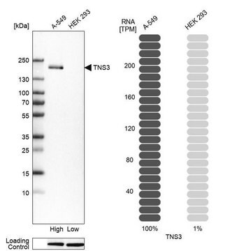
Western Blot Analysis: 1 µg/mL of this antibody detected Tensin-3 in 10 µg of human kidney tissue lysate.

Target description

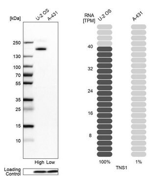
~180 kDa observed.
The calculated molecular weight is 155 kDa, however Tensin-3 has been shown as a ~180 kDa band in western blots (Cui, Y., et al. (2004). Molecular Cancer Research. 2:225–232).
An uncharacterized band appears at ~22 kDa in some lysates.

Physical form

Affinity purified
Purified rabbit polyclonal in buffer containing 0.1 M Tris-Glycine (pH 7.4), 150 mM NaCl with 0.05% sodium azide.

Storage and Stability

Stable for 1 year at 2-8°C from date of receipt.

Analysis Note

Control
Human kidney tissue lysate

Other Notes

Concentration: Please refer to the Certificate of Analysis for the lot-specific concentration.

Disclaimer

Unless otherwise stated in our catalog or other company documentation accompanying the product(s), our products are intended for research use only and are not to be used for any other purpose, which includes but is not limited to, unauthorized commercial uses, in vitro diagnostic uses, ex vivo or in vivo therapeutic uses or any type of consumption or application to humans or animals.

Not finding the right product?  

Try our Product Selector Tool.

Storage Class Code

12 - Non Combustible Liquids

WGK

WGK 1

Flash Point(F)

Not applicable

Flash Point(C)

Not applicable


Regulatory Listings

Regulatory Listings are mainly provided for chemical products. Only limited information can be provided here for non-chemical products. No entry means none of the components are listed. It is the user’s obligation to ensure the safe and legal use of the product.

JAN Code

ABT29:


Certificates of Analysis (COA)

Search for Certificates of Analysis (COA) by entering the products Lot/Batch Number. Lot and Batch Numbers can be found on a product’s label following the words ‘Lot’ or ‘Batch’.

Already Own This Product?

Find documentation for the products that you have recently purchased in the Document Library.

Visit the Document Library

Nuria Barber-Pérez et al.
Journal of cell science, 133(12) (2020-05-13)
Fibrillar adhesions are important structural and adhesive components in fibroblasts, and are required for fibronectin fibrillogenesis. While nascent and focal adhesions are known to respond to mechanical cues, the mechanoresponsive nature of fibrillar adhesions remains unclear. Here, we used ratiometric

Our team of scientists has experience in all areas of research including Life Science, Material Science, Chemical Synthesis, Chromatography, Analytical and many others.

Contact Technical Service