Skip to Content
Merck
All Photos(3)

Key Documents

Safety Information

ABT135

Sigma-Aldrich

Anti-phospho-FAK (Tyr397) Antibody

from rabbit, purified by affinity chromatography

Synonym(s):

Focal adhesion kinase 1, FADK 1, Protein-tyrosine kinase 2, pp125FAK

Sign Into View Organizational & Contract Pricing


About This Item

UNSPSC Code:
12352203
eCl@ss:
32160702
NACRES:
NA.41
clone:
polyclonal
application:
WB
cell based assay
species reactivity:
rat
technique(s):
cell based assay: suitable
western blot: suitable
citations:
7

biological source

rabbit

Quality Level

antibody form

affinity isolated antibody

antibody product type

primary antibodies

clone

polyclonal

purified by

affinity chromatography

species reactivity

rat

species reactivity (predicted by homology)

human (based on 100% sequence homology), mouse (based on 100% sequence homology), Xenopus (based on 100% sequence homology), chicken (based on 100% sequence homology)

technique(s)

cell based assay: suitable
western blot: suitable

NCBI accession no.

UniProt accession no.

shipped in

wet ice

target post-translational modification

phosphorylation (pTyr397)

Gene Information

human ... PTK2(5747)

General description

Focal adhesion kinase (FAK) is a non receptor protein tyrosine kinase discovered as a substrate for Src and a key element in integrin signaling. FAK plays a central role in cell spreading, differentiation, migration, cell death and acceleration of the G1 to S phase transition of the cell cycle. FAK regulation includes phosphorylation at multiple tyrosine and serine residues. Phosphorylation of tyrosine is generally associated with positive regulation and growth promotion; however, dephosphorylation at these sites occurs as cells enter mitosis (M-Phase). In contrast, serine phosphorylation either remains high or is increased as cells enter mitosis and may play a role in focal adhesion disassembly. FAK and its phosphorylation states have been implicated in cancer metastasis, tumor cell survival, and adhesion-independent growth. Recent evidence indicates that elevation of FAK activity in human carcinoma cells is associated with increased invasive potential. A central role in tumor formation and progression suggests that FAK is an attractive target for therapeutic intervention. Activation of FAK by integrin clustering leads to autophosphorylation at Tyr397.

Specificity

This antibody recognizes FAK phosphorylated at Tyr397.

Immunogen

Epitope: Phosphorylated Tyr397
KLH-conjugated linear peptide corresponding to human FAK phosphorylated at Tyr397.

Application

Detect phospho-FAK (Tyr397) using this Anti-phospho-FAK (Tyr397) Antibody validated for use in WB, Cell Function Assay.
Peptide Inhibition Assay Analysis: A 1:1,000 dilution from a representative lot peptide blocked on RAT2 cells transfected with Src.
Research Category
Cell Structure
Research Sub Category
Adhesion (CAMs)

Quality

Evaluated by Western Blot in untransfected and Src-transfected RAT2 cell lysates.

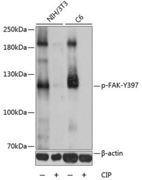
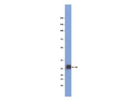
Western Blot Analysis: A 1:1,000 dilution of this antibody detected phospho-FAK (Tyr397) in 10 µg of untransfected and Src-transfected RAT2 cell lysates.

Target description

~125 kDa observed.
An uncharacterized band appears at ~190 kDa in some lysates.

Linkage

Replaces: 04-974

Physical form

Affinity purified
Purified rabbit polyclonal in buffer containing 0.1 M Tris-Glycine (pH 7.4), 150 mM NaCl with 0.05% sodium azide.

Storage and Stability

Stable for 1 year at 2-8°C from date of receipt.

Analysis Note

Control
untransfected and Src-transfected RAT2 cell lysates

Disclaimer

Unless otherwise stated in our catalog or other company documentation accompanying the product(s), our products are intended for research use only and are not to be used for any other purpose, which includes but is not limited to, unauthorized commercial uses, in vitro diagnostic uses, ex vivo or in vivo therapeutic uses or any type of consumption or application to humans or animals.

Not finding the right product?  

Try our Product Selector Tool.

Storage Class Code

12 - Non Combustible Liquids

WGK

WGK 1

Flash Point(F)

Not applicable

Flash Point(C)

Not applicable


Regulatory Listings

Regulatory Listings are mainly provided for chemical products. Only limited information can be provided here for non-chemical products. No entry means none of the components are listed. It is the user’s obligation to ensure the safe and legal use of the product.

JAN Code

ABT135:


Certificates of Analysis (COA)

Search for Certificates of Analysis (COA) by entering the products Lot/Batch Number. Lot and Batch Numbers can be found on a product’s label following the words ‘Lot’ or ‘Batch’.

Already Own This Product?

Find documentation for the products that you have recently purchased in the Document Library.

Visit the Document Library

Jennifer J Chia et al.
The Journal of clinical investigation, 126(11), 4331-4345 (2016-11-02)
Scleroderma is a group of skin-fibrosing diseases for which there are no effective treatments. A feature of the skin fibrosis typical of scleroderma is atrophy of the dermal white adipose tissue (DWAT). Adipose tissue contains adipose-derived mesenchymal stromal cells (ADSCs)
Jae Hoon Kim et al.
International journal of molecular sciences, 23(20) (2022-10-28)
PTK7 is a catalytically defective receptor protein tyrosine kinase upregulated in various cancers, including esophageal squamous cell carcinoma (ESCC). In previous studies, we observed a positive correlation between PTK7 expression levels and tumorigenicity in various ESCC cell lines and xenograft
Defang Pang et al.
Experimental & molecular medicine, 50(9), 117-117 (2018-09-07)
We have previously demonstrated that in response to cerebral ischemia (CI), the growth factor angiopoietin-1 (Ang1) and α5β1 integrin are both induced in cerebral vessels, which likely provide positive signals driving the endogenous angiogenic response and vascular protection after CI.

Our team of scientists has experience in all areas of research including Life Science, Material Science, Chemical Synthesis, Chromatography, Analytical and many others.

Contact Technical Service