Skip to Content
Merck
All Photos(2)

Key Documents

Safety Information

AB1768-I

Sigma-Aldrich

Anti-GluR2 Antibody

from rabbit, purified by affinity chromatography

Synonym(s):

Glutamate receptor 2, GluR-2, AMPA-selective glutamate receptor 2, GluR-B, GluR-K2, Glutamate receptor ionotropic, AMPA 2, GluA2

Sign Into View Organizational & Contract Pricing


About This Item

UNSPSC Code:
12352203
eCl@ss:
32160702
NACRES:
NA.41

biological source

rabbit

Quality Level

antibody form

affinity isolated antibody

antibody product type

primary antibodies

clone

polyclonal

purified by

affinity chromatography

species reactivity

mouse, rat, human

packaging

antibody small pack of 25 μg

technique(s)

immunohistochemistry: suitable (paraffin)
western blot: suitable

NCBI accession no.

UniProt accession no.

shipped in

wet ice

target post-translational modification

unmodified

Gene Information

human ... GRIA2(2891)

General description

Glutamate receptors (GluRs) can be categorized as ionotropic or metabotropic and subcatergorized by their agonist preferences (NMDA, AMPA or Kainic acid). There are four types of AMPA selective GluR subunits (GluR1, GluR2, GluR3 and GluR4). Tetrameric or pentameric combinations of different subunits contributes to the functional diversity of AMPA receptors. In general, AMPA receptors mediate fast synaptic current at most excitatory synapses, with stoichiometry characterized by subtype composition. Although subunit composition of AMPA receptors varies, they must contain at least one edited GluR2 subunit to be calcium impermeable. Relative calcium permeability in AMPA receptor channels may be significant in pathological neurotoxic damage and long term changes in nervous system responses.

Specificity

This antibody recognizes the cytoplasmic domain of Glutamate Receptor 2.

Immunogen

Epitope: Cytoplasmic domain
KLH-conjugated linear peptide corresponding to the cytoplasmic domain of rat GluR2.

Application

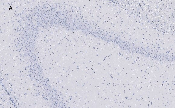
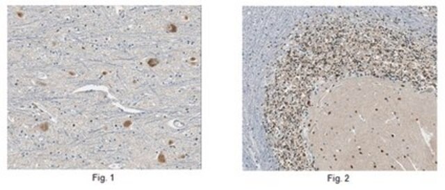
Immunohistochemistry Analysis: A 1:4,000 dilution from a representative lot detected GluR2 in normal human and rat brain tissues.
Research Category
Neuroscience
Research Sub Category
Signaling Neuroscience
This GluR2 antibody is validated for use in WB & IHC for the detection of the GluR2 protein.

Quality

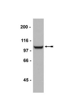
Evaluated by Western Blotting in mouse brain tissue lysate.

Western Blotting Analysis: 0.5 µg/mL of this antibody detected GluR2 in 10 µg of mouse brain tissue lysate.

Target description

~108 kDa observed. Three isoforms at 99 kDa, 99 kDa, and 100 kDa are known to exist due to alternative splicing.
There is no known homology to mouse and rat GluR1, GluR3 and GluR4. There was no homology detected to GluR1 and GluR3 in human, but there is 67% sequence homology to GluR4. An uncharacterized band may be observed at ~75 kDa in some cell lysates.

Linkage

Replaces: AB1768-25UG

Physical form

Affinity purified
Purified rabbit polyclonal in buffer containing 0.1 M Tris-Glycine (pH 7.4), 150 mM NaCl with 0.05% sodium azide.

Storage and Stability

Stable for 1 year at 2-8°C from date of receipt.

Analysis Note

Control
Mouse brain tissue lysate

Other Notes

Concentration: Please refer to the Certificate of Analysis for the lot-specific concentration.

Disclaimer

Unless otherwise stated in our catalog or other company documentation accompanying the product(s), our products are intended for research use only and are not to be used for any other purpose, which includes but is not limited to, unauthorized commercial uses, in vitro diagnostic uses, ex vivo or in vivo therapeutic uses or any type of consumption or application to humans or animals.

Not finding the right product?  

Try our Product Selector Tool.

Storage Class Code

12 - Non Combustible Liquids

WGK

WGK 1

Flash Point(F)

Not applicable

Flash Point(C)

Not applicable


Regulatory Listings

Regulatory Listings are mainly provided for chemical products. Only limited information can be provided here for non-chemical products. No entry means none of the components are listed. It is the user’s obligation to ensure the safe and legal use of the product.

JAN Code

AB1768-I:


Certificates of Analysis (COA)

Search for Certificates of Analysis (COA) by entering the products Lot/Batch Number. Lot and Batch Numbers can be found on a product’s label following the words ‘Lot’ or ‘Batch’.

Already Own This Product?

Find documentation for the products that you have recently purchased in the Document Library.

Visit the Document Library

Meenakshi M Asokan et al.
Nature communications, 9(1), 2468-2468 (2018-06-27)
Layer 5 (L5) cortical projection neurons innervate far-ranging brain areas to coordinate integrative sensory processing and adaptive behaviors. Here, we characterize a plasticity in L5 auditory cortex (ACtx) neurons that innervate the inferior colliculus (IC), thalamus, lateral amygdala and striatum.
Ji-Eun Kim et al.
International journal of molecular sciences, 21(16) (2020-08-13)
α-Amino-3-hydroxy-5-methyl-4-isoxazolepropionic acid receptor (AMPAR) is one of the ligand-gated ion channels for glutamate, which is an important player in the generation and spread of seizures. The efficacy of AMPAR functionality is regulated by the trafficking, synaptic targeting, and phosphorylation. Paradoxically
Lei-Lei Liu et al.
Molecular vision, 25, 780-790 (2019-12-11)
The neuromodulator dopamine plays an important role in light adaptation for the visual system. Light can stimulate dopamine release from dopaminergic amacrine cells (DACs) by activating three classes of photosensitive retinal cells: rods, cones, and melanopsin-expressing intrinsically photosensitive retinal ganglion
Sung Eun Wang et al.
Experimental neurobiology, 28(4), 537-546 (2019-09-09)
Silent information regulator 2 (Sirtuin2 / SIRT2) is a NAD+-dependent deacetylase that regulates the cellular oxidative stress response. It modulates transcriptional silencing and protein stability through deacetylation of target proteins including histones. Previous studies have shown that SIRT2 plays a
Duk-Shin Lee et al.
Scientific reports, 13(1), 13927-13927 (2023-08-26)
Protein disulfide isomerase (PDI) is a redox-active enzyme and also serves as a nitric oxide donor causing S-nitrosylation of cysteine residues in various proteins. Although PDI knockdown reduces α-amino-3-hydroxy-5-methylisoxazole-4-propionic acid receptor (AMPAR)-mediated neuronal activity, the underlying mechanisms are largely unknown.

Our team of scientists has experience in all areas of research including Life Science, Material Science, Chemical Synthesis, Chromatography, Analytical and many others.

Contact Technical Service