Skip to Content
Merck
All Photos(1)

Key Documents

Safety Information

AB1557P

Sigma-Aldrich

Anti-NMDAR2B Antibody

Chemicon®, from rabbit

Synonym(s):

N-methyl D-aspartate receptor subtype 2B, N-methyl-D-aspartate receptor subunit 2B, N-methyl-D-aspartate receptor subunit 3, glutamate receptor subunit epsilon-2, glutamate receptor. ionotropic, N-methyl D-aspartate 2B, Glutamate

Sign Into View Organizational & Contract Pricing


About This Item

UNSPSC Code:
12352203
eCl@ss:
32160702
NACRES:
NA.41

biological source

rabbit

Quality Level

antibody form

affinity isolated antibody

antibody product type

primary antibodies

clone

polyclonal

species reactivity

mouse, rat, human

manufacturer/tradename

Chemicon®

technique(s)

immunohistochemistry: suitable
immunoprecipitation (IP): suitable
western blot: suitable

NCBI accession no.

UniProt accession no.

shipped in

dry ice

target post-translational modification

unmodified

General description

NMDA receptors are a class of ionotropic glutamate receptors. NMDA receptor channel has been shown to be involved in long term potentiation, an activity dependent increase in the efficiency of synaptic transmission thought to underlie certain kinds of memory and learning. NMDA receptor channels are heteromers composed of the key receptor subunit NMDAR1 (GRIN1) and 1 or more of the 4 NMDAR2 subunits: NMDAR2A (GRIN2A), NMDAR2B (GRIN2B), NMDAR2C (GRIN2C), and NMDAR2D (GRIN2D). GRIN2B may be a candidate gene for the neurodegenerative disorder dentato-rubro-pallidoluysian atrophy (DRPLA).
Properties of NMDAR include modulation by glycine, inhibition by Zn2+, voltage dependent Mg2+ blockade and high Ca2+ permeability. The involvement of NMDAR in the CNS has become a focus area for neurodegenerative diseases such as Alzheimer′s disease and also epilepsy and ischemic neuronal cell death.

Specificity

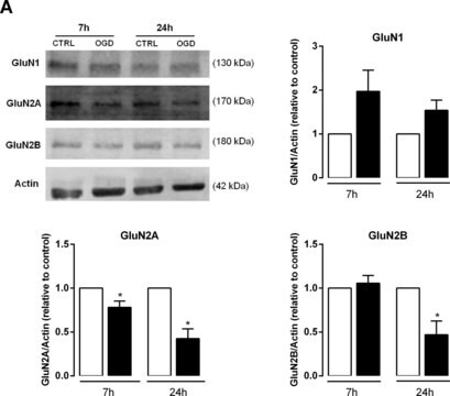
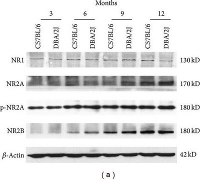
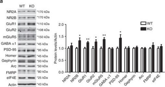
NMDAR2B. Strong and specific immunolabeling of the 180 kDa NMDAR2B subunit of the NMDA receptor. No reactivity to NMDAR2A or NMDAR2C by Western blot. Immunolabeling has been blocked by preadsorption of the antibody with the C-terminal fusion protein that was used to generate the antibody.

Immunogen

C-Terminal Fusion Protein of the NMDAR2B (30 kDa).
Epitope: C-terminus

Application

Anti-NMDAR2B Antibody is an antibody against NMDAR2B for use in IH, IP & WB.
Immunohistochemistry:
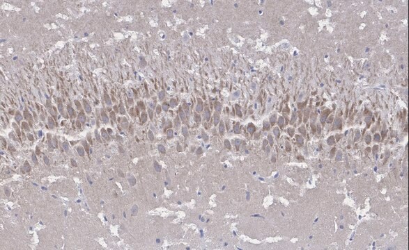
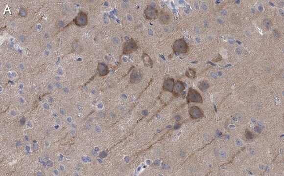
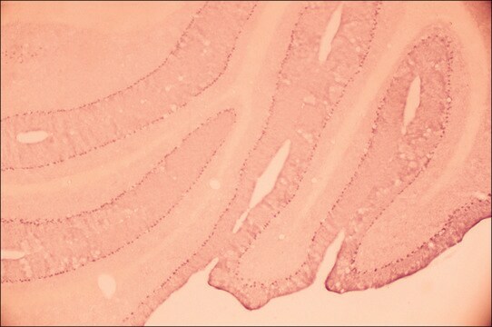
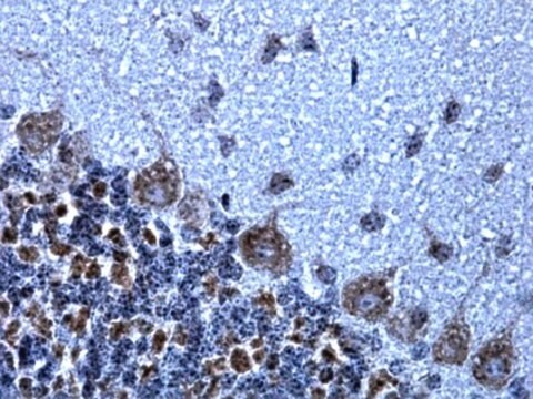
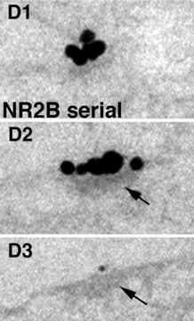
1:1,000-1:2,000 dilution of a previous lot was used on immunohistochemistry.

Immunoprecipitation:
3 μL of a previous lot of this antibody (under appropriate conditions) quantitatively immunoprecipitated all NMDAR2B in 200 μg of rat brain.

Western Blot Analysis:
1:100-1:2,000 dilution of a previous lot (NMDAR2B is present in high concentrations in the hippocampus).

Optimal working dilutions must be determined by the end user.
Research Category
Neuroscience
Research Sub Category
Neurotransmitters & Receptors

Target description

180 kDa

Physical form

Format: Purified
ImmunoAffinity Purified
Purified rabbit polyclonal lyophilized from ammonium bicarbonate (5 mM). Some residual salt may also be present. Reconstitute with 50 μL of PBS. Contains no preservatives.

Storage and Stability

Stable for 1 year at -20ºC from date of receipt.

Analysis Note

Control
Brain tissue, Rat Purkinje cell dendrites, rat hippocampus lysate, HEK 293 cells expressing NR2B.

Other Notes

Concentration: Please refer to the Certificate of Analysis for the lot-specific concentration.

Legal Information

CHEMICON is a registered trademark of Merck KGaA, Darmstadt, Germany

Disclaimer

Unless otherwise stated in our catalog or other company documentation accompanying the product(s), our products are intended for research use only and are not to be used for any other purpose, which includes but is not limited to, unauthorized commercial uses, in vitro diagnostic uses, ex vivo or in vivo therapeutic uses or any type of consumption or application to humans or animals.

Not finding the right product?  

Try our Product Selector Tool.

Storage Class Code

11 - Combustible Solids

WGK

WGK 1


Regulatory Listings

Regulatory Listings are mainly provided for chemical products. Only limited information can be provided here for non-chemical products. No entry means none of the components are listed. It is the user’s obligation to ensure the safe and legal use of the product.

JAN Code

AB1557P:


Certificates of Analysis (COA)

Search for Certificates of Analysis (COA) by entering the products Lot/Batch Number. Lot and Batch Numbers can be found on a product’s label following the words ‘Lot’ or ‘Batch’.

Already Own This Product?

Find documentation for the products that you have recently purchased in the Document Library.

Visit the Document Library

Long-term potentiation in the hippocampal CA1 area and dentate gyrus plays different roles in spatial learning.
Takashi Okada, Nobuaki Yamada, Keisuke Tsuzuki, Hiroshi P M Horikawa, Kohichi Tanaka, Seiji Ozawa
The European Journal of Neuroscience null
Functional NMDA receptor channels generated by NMDAR2B gene transfer in rat cerebellar Purkinje cells.
Wataru Kakegawa, Keisuke Tsuzuki, Masae Iino, Seiji Ozawa
The European Journal of Neuroscience null
Q Liu et al.
Neuroscience, 171(3), 637-654 (2010-10-05)
Previously, we reported that a critical period in respiratory network development exists in rats around postnatal days (P; P12-P13), when abrupt neurochemical, metabolic, and physiological changes occur. Specifically, the expressions of glutamate and N-methyl-d-aspartate (NMDA) receptor (NR) subunit 1 in
A M Khan et al.
The Journal of comparative neurology, 428(3), 428-449 (2000-11-14)
Glutamate (Glu), a major excitatory neurotransmitter within the hypothalamus and thalamus, acts upon many receptors, including the N-methyl-D-aspartate (NMDA) subtype. Abundant evidence suggests that variations in the subunit composition of NMDA receptors (NMDA-Rs) contribute to differences in Glu's immediate electrophysiological
M C Currás-Collazo et al.
The Journal of comparative neurology, 427(1), 93-108 (2000-10-24)
Previous studies suggest that activation of N-methyl-D-aspartate (NMDA) receptors facilitates phasic firing and spike clustering displayed by magnocellular neuroendocrine cells (MNCs) of the supraoptic (SON) and paraventricular nucleus of the hypothalamus (PVN). Osmotic stimulation produces similar activity patterns which, in

Our team of scientists has experience in all areas of research including Life Science, Material Science, Chemical Synthesis, Chromatography, Analytical and many others.

Contact Technical Service