Skip to Content
Merck
All Photos(2)

Key Documents

Safety Information

07-697

Sigma-Aldrich

Anti-E-Cadherin Antibody

serum, Upstate®

Sign Into View Organizational & Contract Pricing


About This Item

UNSPSC Code:
12352203
eCl@ss:
32160702
NACRES:
NA.41

biological source

rabbit

Quality Level

antibody form

serum

antibody product type

primary antibodies

clone

polyclonal

species reactivity

human

packaging

antibody small pack of 25 μL

manufacturer/tradename

Upstate®

technique(s)

immunohistochemistry: suitable
western blot: suitable

isotype

IgG

NCBI accession no.

UniProt accession no.

shipped in

ambient

target post-translational modification

unmodified

Gene Information

human ... CDH1(999)

General description

Cadherin-1 (UniProt: P12830; also known as CAM 120/80, Epithelial cadherin, E-cadherin, Uvomorulin, CD324) is encoded by the CDH1 (also known as CDHE, UVO) gene (Gene ID: 999) in human. Cadherins are calcium-dependent cell adhesion molecules that participate in cell-cell adhesion during embryogenesis, development, organogenesis, and differentiation. E-cadherin is a single-pass type I membrane glycoprotein that is mainly expressed in non-neural epithelial tissues. It is synthesized with a signal peptide (aa 1-22) and a propeptide (aa 23-154) that are subsequently cleaved off to produce the mature form that contains an extracellular domain (aa 155-709), a transmembrane domain (aa 710-730), and a cytoplasmic domain (aa 731-882). N-glycosylation at Asn-637 is shown to be essential for its expression, folding, and trafficking. E-cadherin is known to contain five cadherin domains. Three calcium ions are usually bound at the interface of each cadherin domain and rigidify the connections, imparting a strong curvature to the full-length ectodomain. Post-translationally it can be cleaved into three chains: E-Cad/CTF1 (aa 701-882); E-Cad/CTF2 (aa 732-882); and E-Cad/CTF3 (aa 751-882). During apoptosis or with calcium influx, it is cleaved by a membrane-bound metalloproteinase (ADAM10; at residues 700-701), which causes disruption of cell-cell adhesion and the subsequent release of b-catenin into the cytoplasm. The residual membrane-tethered cleavage product is then rapidly degraded via an intracellular proteolytic pathway. It can also be cleaved by PS1/g-secretase (at residues 731-732) and this cleavage promotes disassembly of adherens junctions. Caspase 3 can cleave it at residues 750-751, which releases the cytoplasmic tail resulting in disintegration of the actin microfilament system. (Ref.: Marambaud, M., et al. (2002). EMBO J. 21(8); 1948-1956; Steinhausen, U., et al. (2001). J. Biol. Chem. 276(7); 4972-4980; Ito, K., et al. (1999). Oncogene. 18(50); 7080-7090).

Specificity

E-Cadherin
Predicted to cross-react with mouse, rat, orangutan, bovine, chicken and canine based on sequence homology

Immunogen

synthetic peptide corresponding to amino acids 859-874 of human E-Cadherin

Application

Detect E-Cadherin using this Anti-E-Cadherin Antibody validated for use in Immunohistochemistry and WB.

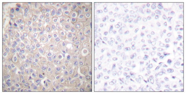
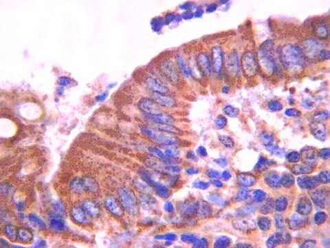
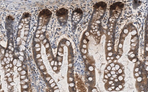
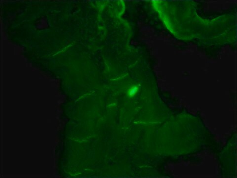
Immunohistochemistry (Paraffin) Analysis: A 1:250 dilution of this antibody detected E-Cadherin in Human small intestine tissue sections.
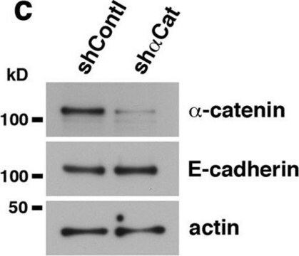
Western Blotting Analysis: A 1:1,000 dilution of this antibody detected E-Cadherin in HepG2 cell lysate.
Research Category
Cell Structure
Research Sub Category
Adhesion (CAMs)

Quality

Routinely evaluated by immunoblot with RIPA lysates from HepG2 cells.

Target description

106kDa

Physical form

Serum

Storage and Stability

2 years at -20°C from date of shipment

Other Notes

Please refer to lot specific datasheet.

Legal Information

UPSTATE is a registered trademark of Merck KGaA, Darmstadt, Germany

Disclaimer

Unless otherwise stated in our catalog or other company documentation accompanying the product(s), our products are intended for research use only and are not to be used for any other purpose, which includes but is not limited to, unauthorized commercial uses, in vitro diagnostic uses, ex vivo or in vivo therapeutic uses or any type of consumption or application to humans or animals.

Not finding the right product?  

Try our Product Selector Tool.

Storage Class Code

10 - Combustible liquids

WGK

WGK 1


Regulatory Listings

Regulatory Listings are mainly provided for chemical products. Only limited information can be provided here for non-chemical products. No entry means none of the components are listed. It is the user’s obligation to ensure the safe and legal use of the product.

JAN Code

07-697BR:
07-697-25UL:
07-697:
07-697-100UL:


Certificates of Analysis (COA)

Search for Certificates of Analysis (COA) by entering the products Lot/Batch Number. Lot and Batch Numbers can be found on a product’s label following the words ‘Lot’ or ‘Batch’.

Already Own This Product?

Find documentation for the products that you have recently purchased in the Document Library.

Visit the Document Library

Sarah Christine Pereira de Oliveira et al.
Molecular nutrition & food research, 65(9), e2000863-e2000863 (2021-03-03)
Nutritional supplementation of the maternal diet can modify the cancer susceptibility in adult offspring. Therefore, the authors evaluate the effects of a fish-oil diet administered to a long-term, during pre-mating, gestation, and lactation, in reducing cancer-cachexia damages in adult Walker-256
Bcl3 selectively promotes metastasis of ERBB2-driven mammary tumors.
Wakefield, A; Soukupova, J; Montagne, A; Ranger, J; French, R; Muller, WJ; Clarkson, RW
Cancer Research null
Tight and adherens junctions in the ovine uterus: differential regulation by pregnancy and progesterone.
Satterfield, MC; Dunlap, KA; Hayashi, K; Burghardt, RC; Spencer, TE; Bazer, FW
Endocrinology null
Yu-Jen Chiu et al.
International journal of oncology, 52(5), 1465-1478 (2018-03-24)
Osteosarcoma is the most common primary malignancy of the bone and is characterized by local invasion and distant metastasis. Over the past 20 years, long-term outcomes have reached a plateau even with aggressive therapy. Overexpression of insulin-like growth factor 1 receptor
Direct transcriptional activation of promyelocytic leukemia protein by IFN regulatory factor 3 induces the p53-dependent growth inhibition of cancer cells.
Kim, TK; Lee, JS; Oh, SY; Jin, X; Choi, YJ; Lee, TH; Lee, Eh; Choi, YK; You, S; Chung et al.
Cancer Research null

Our team of scientists has experience in all areas of research including Life Science, Material Science, Chemical Synthesis, Chromatography, Analytical and many others.

Contact Technical Service