Skip to Content
Merck
All Photos(1)

Key Documents

Safety Information

07-326

Sigma-Aldrich

Anti-phospho-Cofilin 2 (Ser3) Antibody

Upstate®, from rabbit

Sign Into View Organizational & Contract Pricing


About This Item

UNSPSC Code:
12352203
eCl@ss:
32160702
NACRES:
NA.41

biological source

rabbit

Quality Level

antibody form

purified immunoglobulin

antibody product type

primary antibodies

clone

polyclonal

species reactivity

mouse, chicken, human

manufacturer/tradename

Upstate®

technique(s)

western blot: suitable

isotype

IgG

NCBI accession no.

UniProt accession no.

shipped in

dry ice

target post-translational modification

phosphorylation (pSer3)

Gene Information

human ... CFL2(1073)

Specificity

phospho-Cofilin 2

Immunogen

peptide corresponding to amino acids 1-10 (MA[pS]GVTVNDE-C) of human phospho-Cofilin 2

Application

Anti-phospho-Cofilin 2 (Ser3) Antibody detects level of phospho-Cofilin 2 (Ser3) & has been published & validated for use in WB.
Research Category
Cell Structure
Research Sub Category
Cytoskeleton

Quality

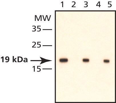
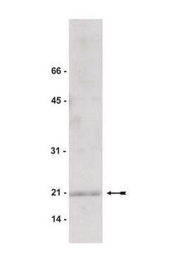
routinely evaluated by immunoblot on RIPA lysates from L6 cells, treated with LIM Kinase

Target description

21kDa

Physical form

0.1M Tris-glycine, pH 7.4, 0.15M NaCl, 0.05% sodium azide before the addition of glycerol to 30%
Format: Purified
Protein A chromatography

Storage and Stability

2 years at -20°C

Legal Information

UPSTATE is a registered trademark of Merck KGaA, Darmstadt, Germany

Disclaimer

Unless otherwise stated in our catalog or other company documentation accompanying the product(s), our products are intended for research use only and are not to be used for any other purpose, which includes but is not limited to, unauthorized commercial uses, in vitro diagnostic uses, ex vivo or in vivo therapeutic uses or any type of consumption or application to humans or animals.

Not finding the right product?  

Try our Product Selector Tool.

Storage Class Code

10 - Combustible liquids

WGK

WGK 1


Regulatory Listings

Regulatory Listings are mainly provided for chemical products. Only limited information can be provided here for non-chemical products. No entry means none of the components are listed. It is the user’s obligation to ensure the safe and legal use of the product.

JAN Code

07-326:


Certificates of Analysis (COA)

Search for Certificates of Analysis (COA) by entering the products Lot/Batch Number. Lot and Batch Numbers can be found on a product’s label following the words ‘Lot’ or ‘Batch’.

Already Own This Product?

Find documentation for the products that you have recently purchased in the Document Library.

Visit the Document Library

C Thirion et al.
European journal of biochemistry, 268(12), 3473-3482 (2001-06-26)
Cofilins are actin binding proteins and regulate actin assembly in vivo. Numerous cofilin homologues have been characterized in various organisms including mammals. In mice, a ubiquitously expressed cofilin (CFL1) and a skeletal muscle specific cofilin (CFL2) have been described. In
J Craig Hunter et al.
Journal of molecular and cellular cardiology, 47(6), 810-818 (2009-10-06)
Although nitric oxide (NO) has received extensive attention as an anti-hypertrophic agent the mechanisms underlying its regulation of endothelin-1 (ET-1) have not been fully elucidated. Since RhoA has been identified as an important mediator of cardiac hypertrophy and is inhibited
Differential dependence of stretch and shear stress signaling on caveolin-1 in the vascular wall.
Albinsson, S; Nordstrom, I; Sward, K; Hellstrand, P
American Journal of Physiology. Cell Physiology null
Toru Amano et al.
The Journal of biological chemistry, 277(24), 22093-22102 (2002-04-02)
Actin filament dynamics play a critical role in mitosis and cytokinesis. LIM motif-containing protein kinase 1 (LIMK1) regulates actin reorganization by phosphorylating and inactivating cofilin, an actin-depolymerizing and -severing protein. To examine the role of LIMK1 and cofilin during the
Mardjaneh Sadegh Karbalaei et al.
European journal of pharmacology, 689(1-3), 179-185 (2012-05-31)
Caveolae are membrane invaginations present in a variety of cell types. Formation of caveolae depends on caveolins and on the more recently discovered family of proteins known as the cavins. Genetic ablation of caveolin-1 was previously shown to give rise

Our team of scientists has experience in all areas of research including Life Science, Material Science, Chemical Synthesis, Chromatography, Analytical and many others.

Contact Technical Service