Passa al contenuto
Merck
Tutte le immagini(2)

Key Documents

P1093

Sigma-Aldrich

Monoclonal Anti-Paxillin antibody produced in mouse

clone PXC-10, ascites fluid

Autenticatiper visualizzare i prezzi riservati alla tua organizzazione & contrattuali


About This Item

Numero MDL:
Codice UNSPSC:
12352203
NACRES:
NA.41

Origine biologica

mouse

Coniugato

unconjugated

Forma dell’anticorpo

ascites fluid

Tipo di anticorpo

primary antibodies

Clone

PXC-10, monoclonal

PM

antigen 68-40 kDa

contiene

15 mM sodium azide

Reattività contro le specie

bovine, rat, human, chicken, hamster

tecniche

immunocytochemistry: suitable
microarray: suitable
western blot: 1:500 using a cultured human cell line extract

Isotipo

IgG1

N° accesso UniProt

Condizioni di spedizione

dry ice

Temperatura di conservazione

−20°C

modifica post-traduzionali bersaglio

unmodified

Informazioni sul gene

human ... PXN(5829)
rat ... Pxn(360820)

Descrizione generale

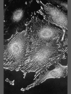
Monoclonal Anti-Paxillin (mouse IgG1 isotype) is derived from the PXC-10 hybridoma produced by the fusion of mouse myeloma cells and splenocytes from an immunized mouse. Paxillin (68 kDa), is a cytoskeletal component. It is found in the ends of actin stress fibers of the focal adhesions, but not in adherens junctions of the cells. The paxillin molecule has a single binding site for vinculin, and at least two binding sites for focal adhesion kinase (FAK), that are separated by an intervening sequence of 100 amino acids.

Specificità

Recognizes an epitope located in the LIM1 domain of the paxillin molecule.

Immunogeno

C-terminal part of recombinant chicken paxillin (amino acids 305-559)

Applicazioni

Monoclonal Anti-Paxillin antibody produced in mouse has been used in immunoblotting and immunocytochemistry.

Azioni biochim/fisiol

Paxillin interacts with several proteins including members of the src family of tyrosine kinases, the transforming protein v-crk, the cytoskeletal protein vinculin, and the tyrosine kinase, focal adhesion kinase (FAK). This interaction has suggested a function for paxillin as a molecular adaptor, responsible for the recruitment of structural and signaling molecules to focal adhesions.

Esclusione di responsabilità

Unless otherwise stated in our catalog or other company documentation accompanying the product(s), our products are intended for research use only and are not to be used for any other purpose, which includes but is not limited to, unauthorized commercial uses, in vitro diagnostic uses, ex vivo or in vivo therapeutic uses or any type of consumption or application to humans or animals.

Non trovi il prodotto giusto?  

Prova il nostro Motore di ricerca dei prodotti.

Codice della classe di stoccaggio

10 - Combustible liquids

Classe di pericolosità dell'acqua (WGK)

WGK 3

Punto d’infiammabilità (°F)

Not applicable

Punto d’infiammabilità (°C)

Not applicable


Certificati d'analisi (COA)

Cerca il Certificati d'analisi (COA) digitando il numero di lotto/batch corrispondente. I numeri di lotto o di batch sono stampati sull'etichetta dei prodotti dopo la parola ‘Lotto’ o ‘Batch’.

Possiedi già questo prodotto?

I documenti relativi ai prodotti acquistati recentemente sono disponibili nell’Archivio dei documenti.

Visita l’Archivio dei documenti

Paxillin: a new vinculin-binding protein present in focal adhesions.
Turner CE, et al.
The Journal of cell biology, 111(3), 1059-1068 (1990)
Paxillin: a crossroad in pathological cell migration
Lopez CAM, et al.
Journal of Hematology & Oncology, 10(1), 50-50 (2017)
Jung Yul Lim et al.
Biomaterials, 28(10), 1787-1797 (2007-01-16)
An important consideration in developing physical biomimetic cell-stimulating cues is that the in vivo extracellular milieu includes nanoscale topographic interfaces. We investigated nanoscale topography regulation of cell functions using human fetal osteoblastic (hFOB) cell culture on poly(l-lactic acid) and polystyrene
Gokhan Bahcecioglu et al.
International journal of biological macromolecules, 122, 1152-1162 (2018-09-16)
In this study, porcine fibrochondrocyte-seeded agarose, methacrylated gelatin (GelMA), methacrylated hyaluronic acid (MeHA) and GelMA-MeHA blend hydrogels, and 3D printed PCL scaffolds were tested under dynamic compression for potential meniscal regeneration in vitro. Cell-carrying hydrogels produced higher levels of extracellular
J L Funderburgh et al.
The Journal of biological chemistry, 276(47), 44173-44178 (2001-09-14)
Keratocytes of the corneal stroma secrete a unique population of proteoglycan molecules considered essential for corneal transparency. In healing corneal wounds, keratocytes exhibit a myofibroblastic phenotype in response to transforming growth factor beta (TGF-beta), characterized by expression of alpha-smooth muscle

Il team dei nostri ricercatori vanta grande esperienza in tutte le aree della ricerca quali Life Science, scienza dei materiali, sintesi chimica, cromatografia, discipline analitiche, ecc..

Contatta l'Assistenza Tecnica.