Passa al contenuto
Merck
Tutte le immagini(5)

Documenti fondamentali

HPA010734

Sigma-Aldrich

Anti-CD1A antibody produced in rabbit

enhanced validation

Prestige Antibodies® Powered by Atlas Antibodies, affinity isolated antibody, buffered aqueous glycerol solution

Sinonimo/i:

Anti-CD1a antigen antibody produced in rabbit, Anti-T-cell surface antigen T6/Leu-6 antibody produced in rabbit, Anti-T-cell surface glycoprotein CD1a precursor antibody produced in rabbit, Anti-hTa1 thymocyte antigen antibody produced in rabbit

Autenticatiper visualizzare i prezzi riservati alla tua organizzazione & contrattuali


About This Item

Numero MDL:
Codice UNSPSC:
12352203
Numero Human Protein Atlas:
NACRES:
NA.41

Origine biologica

rabbit

Livello qualitativo

Coniugato

unconjugated

Forma dell’anticorpo

affinity isolated antibody

Tipo di anticorpo

primary antibodies

Clone

polyclonal

Nome Commerciale

Prestige Antibodies® Powered by Atlas Antibodies

Stato

buffered aqueous glycerol solution

Reattività contro le specie

human

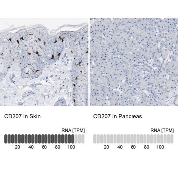
Convalida avanzata

orthogonal RNAseq
Learn more about Antibody Enhanced Validation

tecniche

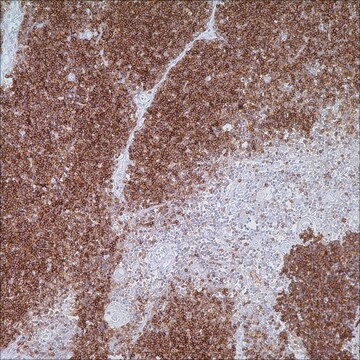
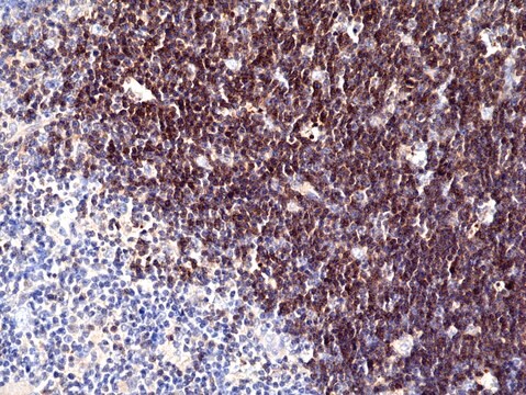
immunohistochemistry: 1:50- 1:200

Sequenza immunogenica

QNLVSGWLSDLQTHTWDSNSSTIVFLCPWSRGNFSNEEWKELETLFRIRTIRSFEGIRRYAHELQFEYPFEIQVTGGCELHSGKVSGSFLQLAYQGSDFVSFQNNSWLPYPVAGNMAKHFCKVLNQNQHEND

N° accesso UniProt

applicazioni

research pathology

Condizioni di spedizione

wet ice

Temperatura di conservazione

−20°C

modifica post-traduzionali bersaglio

unmodified

Informazioni sul gene

human ... CD1A(909)

Descrizione generale

CD1A (cluster of differentiation 1A) belongs to a family of antigen-presenting proteins called CD1. These are evolutionary conserved proteins which present lipids instead of peptide. Humans contain five CD1 genes namely, CD1A, CD1B, CD1C, CD1D and CD1E. CD1A is a transmembrane glycoprotein which is expressed on dendritic cells. It is the only isoform, in this family, to be expressed by Langerhans cells.

Immunogeno

T-cell surface glycoprotein CD1a precursor recombinant protein epitope signature tag (PrEST)

Applicazioni

All Prestige Antibodies Powered by Atlas Antibodies are developed and validated by the Human Protein Atlas (HPA) project and as a result, are supported by the most extensive characterization in the industry.

The Human Protein Atlas project can be subdivided into three efforts: Human Tissue Atlas, Cancer Atlas, and Human Cell Atlas. The antibodies that have been generated in support of the Tissue and Cancer Atlas projects have been tested by immunohistochemistry against hundreds of normal and disease tissues and through the recent efforts of the Human Cell Atlas project, many have been characterized by immunofluorescence to map the human proteome not only at the tissue level but now at the subcellular level. These images and the collection of this vast data set can be viewed on the Human Protein Atlas (HPA) site by clicking on the Image Gallery link. We also provide Prestige Antibodies® protocols and other useful information.

Azioni biochim/fisiol

CD1 (cluster of differentiation 1) family of proteins is responsible for the capture and presentation of self and microbial glycolipids to the specific T cells. CD1A plays an important role in the host defense mechanism against microbial infection. It also facilitates early stages of immune response through CD1A-reactive T cells. This is achieved by inducing cytokine production and dendritic cell maturation, and activating cells such as NK (natural killer) cells and macrophages. The SNP rs411089, present in an intron in the CD1A gene is associated with increased susceptibility to tuberculosis, in Vietnamese population. Hence, it can be hypothesized that CD1A plays an important role in host defense against tuberculosis. The expression of this protein is also increased in monocyte culture derived from untreated multiple sclerosis (MS) patients, as opposed to expression in monocyte culture obtained from treated MS patients.

Caratteristiche e vantaggi

Prestige Antibodies® are highly characterized and extensively validated antibodies with the added benefit of all available characterization data for each target being accessible via the Human Protein Atlas portal linked just below the product name at the top of this page. The uniqueness and low cross-reactivity of the Prestige Antibodies® to other proteins are due to a thorough selection of antigen regions, affinity purification, and stringent selection. Prestige antigen controls are available for every corresponding Prestige Antibody and can be found in the linkage section.

Every Prestige Antibody is tested in the following ways:
  • IHC tissue array of 44 normal human tissues and 20 of the most common cancer type tissues.
  • Protein array of 364 human recombinant protein fragments.

Linkage

Corresponding Antigen APREST72090

Stato fisico

Solution in phosphate-buffered saline, pH 7.2, containing 40% glycerol and 0.02% sodium azide

Note legali

Prestige Antibodies is a registered trademark of Merck KGaA, Darmstadt, Germany

Esclusione di responsabilità

Unless otherwise stated in our catalog or other company documentation accompanying the product(s), our products are intended for research use only and are not to be used for any other purpose, which includes but is not limited to, unauthorized commercial uses, in vitro diagnostic uses, ex vivo or in vivo therapeutic uses or any type of consumption or application to humans or animals.

Non trovi il prodotto giusto?  

Prova il nostro Motore di ricerca dei prodotti.

Codice della classe di stoccaggio

10 - Combustible liquids

Classe di pericolosità dell'acqua (WGK)

WGK 1

Punto d’infiammabilità (°F)

Not applicable

Punto d’infiammabilità (°C)

Not applicable

Dispositivi di protezione individuale

Eyeshields, Gloves, multi-purpose combination respirator cartridge (US)


Scegli una delle versioni più recenti:

Certificati d'analisi (COA)

Lot/Batch Number

Non trovi la versione di tuo interesse?

Se hai bisogno di una versione specifica, puoi cercare il certificato tramite il numero di lotto.

Possiedi già questo prodotto?

I documenti relativi ai prodotti acquistati recentemente sono disponibili nell’Archivio dei documenti.

Visita l’Archivio dei documenti

J Salamero et al.
The Journal of investigative dermatology, 116(3), 401-408 (2001-03-07)
In this work, we studied the localization and traffic of CD1a molecules in human epidermal Langerhans cells and the ability of these cells to stimulate CD1a-restricted T cell clones. We found that CD1a was spontaneously internalized into freshly isolated Langerhans
C Günther et al.
Clinical and experimental dermatology, 37(2), 169-176 (2011-12-23)
The immune system of the skin has a network of resident dendritic cells (DCs) consisting of epidermal Langerhans cells and various subsets of dermal DCs. We recently reported on a new population of dermal DCs, called slan (6-sulfoLacNAc+) DCs, which
C Seshadri et al.
Genes and immunity, 15(3), 195-198 (2014-02-07)
CD1 proteins are antigen-presenting molecules that evolved to present lipids rather than peptides to T cells. However, unlike major histocompatibility complex genes, CD1 genes show low rates of polymorphism and have not been clearly associated with human disease. We report
Claudia Günther et al.
The American journal of pathology, 179(3), 1434-1442 (2011-07-12)
Mycosis fungoides (MF) is the most frequent form of cutaneous T-cell lymphoma (CTCL), which can deteriorate from patch stage to dermal-based tumors and systemic involvement in years. The interaction of chemokines in the skin with CTCL cells might have implications
S Bine et al.
Clinical and experimental immunology, 169(1), 10-16 (2012-06-08)
Studies of multiple sclerosis (MS) have concentrated mainly on antigen presentation of peptides derived from the myelin sheath, while the implication of lipid antigen has been less explored in this pathology. As the extracellular environment regulates expression of the lipid

Il team dei nostri ricercatori vanta grande esperienza in tutte le aree della ricerca quali Life Science, scienza dei materiali, sintesi chimica, cromatografia, discipline analitiche, ecc..

Contatta l'Assistenza Tecnica.