
MABT818
Anti-Myosin-3 (MYH3) Antibody, clone BF-G6
clone BF-G6, from mouse
Sinonimo/i:
Myosin-3, Muscle embryonic myosin heavy chain, Myosin heavy chain 3, Myosin heavy chain, fast skeletal muscle, embryonic, SMHCE
About This Item
Prodotti consigliati

Origine biologica
mouse
Livello qualitativo
Forma dell’anticorpo
purified immunoglobulin
Tipo di anticorpo
primary antibodies
Clone
BF-G6, monoclonal
Reattività contro le specie
rat, human
Reattività contro le specie (prevista in base all’omologia)
bovine (based on 100% sequence homology)
tecniche
ELISA: suitable
immunocytochemistry: suitable
immunofluorescence: suitable
immunohistochemistry: suitable
western blot: suitable
Isotipo
IgG2b, kappa
N° accesso NCBI
N° accesso UniProt
Condizioni di spedizione
ambient
modifica post-traduzionali bersaglio
unmodified
Informazioni sul gene
human ... MYH3(4621)
Descrizione generale
Specificità
Immunogeno
Applicazioni
Cell Structure
ELISA Analysis: Clone BF-G6 hybridoma culture supernatant detected myosin from 10-week human fetus, but not myosin from 8-day new born muscle or adult skeletal muscle (Schiaffino, S., et al. (1986). Exp. Cell Res. 163(1):211-220).
Immunocytochemistry Analysis: Clone BF-G6 hybridoma culture supernatant immunostained aceton-fixed tumor cells in bone marrow aspiration from a child with rhobdomyosarcoma (Schiaffino, S., et al. (1986). Exp. Cell Res. 163(1):211-220).
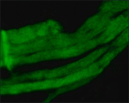
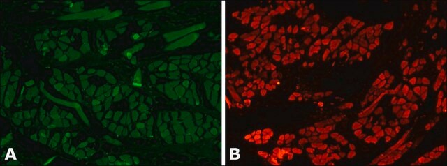
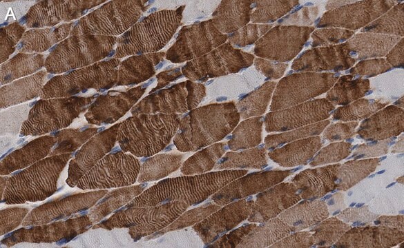
Immunofluorescence Analysis: Clone BF-G6 hybridoma culture supernatant immunostained serial transverse cryosections of rat extraocular (EO) and soleus muscles. BF-G6 staining pattern is similar, but not identical to that of MYH15 (Rossi, A.C., et al. (2010). J. Physiol. 588(Pt 2):353-364).
Immunofluorescence Analysis: Clone BF-G6 hybridoma culture supernatant immunostained frozen fetal muscle sections, while very few cells were stained in 8-week new born muscle tissue and no staining of 8-month infant muscle tissue was observed (Schiaffino, S., et al. (1986). Exp. Cell Res. 163(1):211-220).
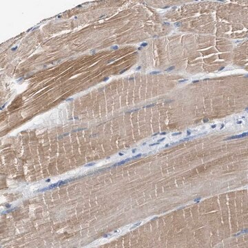
Immunohistochemistry Analysis: Clone BF-G6 hybridoma culture supernatant immunostained tumor cells in frozen human rhobdomyosarcoma sections (Schiaffino, S., et al. (1986). Exp. Cell Res. 163(1):211-220).
Western Blotting Analysis: Clone BF-G6 hybridoma culture supernatant detected myosin from 10-week human fetus, but not myosin from adult skeletal muscle (Schiaffino, S., et al. (1986). Exp. Cell Res. 163(1):211-220).
Qualità
Isotyping Analysis: The identity of this monoclonal antibody is confirmed by isotyping test to be mouse IgG2b, kappa.
Descrizione del bersaglio
Stato fisico
Stoccaggio e stabilità
Altre note
Esclusione di responsabilità
Non trovi il prodotto giusto?
Prova il nostro Motore di ricerca dei prodotti.
Codice della classe di stoccaggio
12 - Non Combustible Liquids
Classe di pericolosità dell'acqua (WGK)
WGK 1
Punto d’infiammabilità (°F)
Not applicable
Punto d’infiammabilità (°C)
Not applicable
Certificati d'analisi (COA)
Cerca il Certificati d'analisi (COA) digitando il numero di lotto/batch corrispondente. I numeri di lotto o di batch sono stampati sull'etichetta dei prodotti dopo la parola ‘Lotto’ o ‘Batch’.
Possiedi già questo prodotto?
I documenti relativi ai prodotti acquistati recentemente sono disponibili nell’Archivio dei documenti.
Il team dei nostri ricercatori vanta grande esperienza in tutte le aree della ricerca quali Life Science, scienza dei materiali, sintesi chimica, cromatografia, discipline analitiche, ecc..
Contatta l'Assistenza Tecnica.