Passa al contenuto
Merck
Tutte le immagini(2)

Key Documents

MABN1775

Sigma-Aldrich

Anti-ABCA4 Antibody, clone TMR1

clone TMR1, from mouse

Sinonimo/i:

Retinal-specific ATP-binding cassette transporter, ATP-binding cassette sub-family A member 4, RIM ABC transporter, RIM protein, RmP, Stargardt disease protein

Autenticatiper visualizzare i prezzi riservati alla tua organizzazione & contrattuali


About This Item

Codice UNSPSC:
12352203
eCl@ss:
32160702

Origine biologica

mouse

Livello qualitativo

Forma dell’anticorpo

purified antibody

Tipo di anticorpo

primary antibodies

Clone

TMR1, monoclonal

Reattività contro le specie

human, mouse

Reattività contro le specie (prevista in base all’omologia)

bovine (based on 100% sequence homology)

tecniche

immunocytochemistry: suitable
immunofluorescence: suitable
western blot: suitable

Isotipo

IgG1κ

N° accesso NCBI

N° accesso UniProt

Condizioni di spedizione

wet ice

modifica post-traduzionali bersaglio

unmodified

Informazioni sul gene

human ... ABCA4(24)

Descrizione generale

Retinal-specific ATP-binding cassette transporter (UniProt F1MWM0/P78363; also known as ATP-binding cassette sub-family A member 4, ABC transporter A4, RIM ABC transporter, RIM protein, RmP, Stargardt disease protein) is encoded by the ABCA4 (also known as ABCR) gene (Gene ID 281584/24) in bovine/human species. ABC transporter A4, a ~250 kDa single-chain, is a multi-pass membrane protein of the superfamily of ATP-binding cassette (ABC) transporters that is localized to the disk margins of vertebrate photoreceptor outer segments. It is implicated in the clearance of all-trans retinal derivatives from rod and cone photoreceptor cells following photoexcitation as part of the visual cycle. ABC transporter A4 is composed of two symmetrically organized halves, each comprising six membrane-spanning helices, a large glycosylated exocytoplasmic domain located inside the disk, and a cytoplasmic domain with an ATP-binding cassette. Mutations in ABCA4 are known to cause Stargardt macular degeneration, which is an autosomal recessive form of juvenile macular degeneration associated with a severe loss in vision.

Ref.: Tsybovsky, Y et al. (2010). Adv. Exp. Med. Biol. 703, 105-125.

Specificità

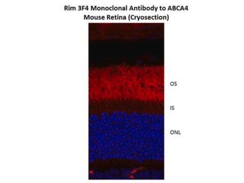
Clone TMR1 immunostained retina cryosections from wild-type mice, but not retina from mice with homozygous L541P;A1038V (PV) missense mutation (Courtesy of Grace Jin, Case Western University, U.S.A.).

Immunogeno

ABCA4 isolated from bovine retinas (Zhang, N., et al. (2015). Hum. Mol. Genet. 24(11):3220-3237).

Applicazioni

Detect ABCA4 using this mouse monoclonal Anti-ABCA4 Antibody, clone TMR1, Cat. No. MABN1775, validated for use in Immunocytochemistry, Immunofluorescence, and Western Blotting.
Immunofluorescence Analysis: A 1:25-125 dilution from a representative lot detected ABCA4 immunoreactivity in retina cryosections from wild-type mice, but not in retina from mice with homozygous L541P;A1038V (PV) missense mutation (Courtesy of Grace Jin, Case Western University, U.S.A.).

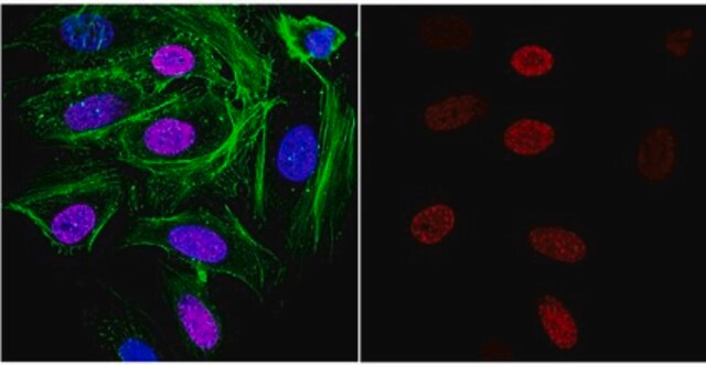
Immunocytochemistry Analysis: A representative lot detected similar cellular localization of wild-type and ABCA4 missense constructs with either or both L541P and A1038V mutation by fluorescent immunocytochemistry staining of 4% paraformaldehyde-fixed, 0.1% Triton X-100-permeabilized HEK293 transfectants (Zhang, N., et al. (2015). Hum. Mol. Genet. 24(11):3220-3237).

Immunofluorescence Analysis: A representative lot detected ABCA4 immunoreactivity in retina cryosections from wild-type mice and mice with heterozygous L541P;A1038V (PV) missense mutaion, but not mice with homozygous PV mutation (Zhang, N., et al. (2015). Hum. Mol. Genet. 24(11):3220-3237).
Research Category
Neuroscience

Qualità

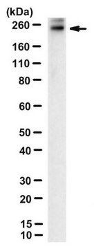
Evaluated by Western Blotting in human retina tissue lysate.

Western Blotting Analysis: A 1:125 dilution of this antibody detected ABCA4 in 10 µg of human retina tissue lysate.


Descrizione del bersaglio

~257 kDa observed. 257.4/255.9/260.2 kDa (bovine/human/mouse) calculated. Uncharacterized bands may be observed in some lysate(s).

Stato fisico

Format: Purified
Protein G purified.
Purified mouse IgG1 in buffer containing 0.1 M Tris-Glycine (pH 7.4), 150 mM NaCl with 0.05% sodium azide.

Stoccaggio e stabilità

Stable for 1 year at 2-8°C from date of receipt.

Altre note

Concentration: Please refer to lot specific datasheet.

Esclusione di responsabilità

Unless otherwise stated in our catalog or other company documentation accompanying the product(s), our products are intended for research use only and are not to be used for any other purpose, which includes but is not limited to, unauthorized commercial uses, in vitro diagnostic uses, ex vivo or in vivo therapeutic uses or any type of consumption or application to humans or animals.

Non trovi il prodotto giusto?  

Prova il nostro Motore di ricerca dei prodotti.

Codice della classe di stoccaggio

12 - Non Combustible Liquids

Classe di pericolosità dell'acqua (WGK)

WGK 1

Punto d’infiammabilità (°F)

Not applicable

Punto d’infiammabilità (°C)

Not applicable


Certificati d'analisi (COA)

Cerca il Certificati d'analisi (COA) digitando il numero di lotto/batch corrispondente. I numeri di lotto o di batch sono stampati sull'etichetta dei prodotti dopo la parola ‘Lotto’ o ‘Batch’.

Possiedi già questo prodotto?

I documenti relativi ai prodotti acquistati recentemente sono disponibili nell’Archivio dei documenti.

Visita l’Archivio dei documenti

Il team dei nostri ricercatori vanta grande esperienza in tutte le aree della ricerca quali Life Science, scienza dei materiali, sintesi chimica, cromatografia, discipline analitiche, ecc..

Contatta l'Assistenza Tecnica.