Passa al contenuto
Merck
Tutte le immagini(2)

Documenti fondamentali

MABF2159

Sigma-Aldrich

Anti-CD63 (LAMP3) Antibody, clone ME491

clone ME491, from mouse

Sinonimo/i:

CD63 antigen, Granulophysin, Lysosomal-associated membrane protein 3, LAMP-3, Melanoma-associated antigen ME491, OMA81H, Ocular melanoma-associated antigen, Tetraspanin-30, Tspan-30

Autenticatiper visualizzare i prezzi riservati alla tua organizzazione & contrattuali


About This Item

Codice UNSPSC:
12352203
eCl@ss:
32160702
NACRES:
NA.43

Origine biologica

mouse

Forma dell’anticorpo

purified immunoglobulin

Tipo di anticorpo

primary antibodies

Clone

ME491, monoclonal

Reattività contro le specie

human

Confezionamento

antibody small pack of 25 μg

tecniche

flow cytometry: suitable
immunocytochemistry: suitable
immunohistochemistry: suitable (paraffin)
western blot: suitable

Isotipo

IgG1κ

N° accesso NCBI

N° accesso UniProt

modifica post-traduzionali bersaglio

unmodified

Informazioni sul gene

human ... CD63(967)

Descrizione generale

CD63 antigen (UniProt: P08962; also known as Granulophysin, Lysosomal-associated membrane protein 3, LAMP-3, Melanoma-associated antigen ME491, OMA81H, Ocular melanoma-associated antigen, Tetraspanin-30, Tspan-30, CD63) is encoded by the CD63 (also known as MLA1, TSPAN30) gene (Gene ID: 967) in human. CD63 is a multi-pass membrane protein of the tetraspan family that is found on endosome, lysosome, and plasma membranes. CD63 has been detected in platelets, Dysplastic nevi benign moles), radial growth phase primary melanomas, hematopoietic cells, and in tissue macrophages. In melanoma cells it is involved in their motility and adhesion. CD63 also plays a role in the adhesion of leukocytes onto endothelial cells. It is reported to play a role in the activation of ITGB1 and integrin signaling, leading to the activation of AKT, FAK/PTK2 and MAP kinases and promote cell survival, reorganization of the actin cytoskeleton, cell adhesion, spreading and migration. CD63 is a highly N-glycosylated protein with three asparagine glycosylation sites (aa 130, 150, 172) and its ribophorin II (RPN2)-mediated glycosylation has been linked to breast cancer. Overexpression of CD63 has been observed in esophageal cancer that is negatively correlated with tumor stage and lymph node metastasis. Lack of expression of CD63 in platelets has been observed in a patient with Hermansky-Pudlak syndrome (HPS), an autosomal recessive disorder that is characterized by oculocutaneous albinism, bleeding due to platelet storage pool deficiency, and lysosomal storage defects. This antibody (clone ME491) is shown to react with human primary and to some extent with metastatic melanoma tissues. (Ref.: Lai, X., et al. (2017). Oncol. Let. 13(6); 4245-4251; Tominaga, N., et al. (2014). Mol. Cancer 13; 134; Smith, M., et al. (1997). Melanoma Res. 7 (Suppl. 2), 163-170).

Specificità

Clone ME491 specifically detects CD63 (LAMP-3) in human cells.

Immunogeno

Clear supernatant from SK-Mel-23 cell lysate.

Applicazioni

Anti-CD63 (LAMP3), clone ME491, Cat. No. MABF2159, is a mouse monoclonal antibody that detects CD63 antigen and has been tested for use in Flow Cytometry, Immunocytochemistry, Immunohistochemistry (Paraffin), and Western Blotting.
Immunohistochemistry (Paraffin) Analysis: A 1:250 dilution from a representative lot detected CD63 (LAMP3) in human spleen and human bone marrow tissue sections.

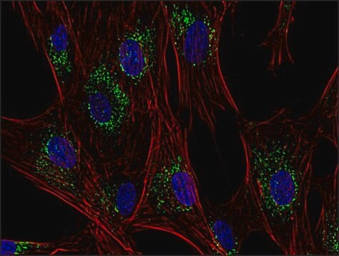
Immunocytochemistry Analysis: A representative lot detected CD63 (LAMP3) in Immunocytochemistry applications (Atkinson, B., et. al. (1984). Cancer Res. 44(6):2577-81).

Flow Cytometry Analysis: A representative lot detected CD63 (LAMP3) in Flow Cytometry applications (Li, J., et. al. (2003). J Immunol. 171(6):2922-9).

Western Blotting Analysis: A representative lot detected CD63 (LAMP3) in Western Blotting applications (Smith, M., et. al. (1997). Melanoma Res. 7 Suppl 2:S163-70).

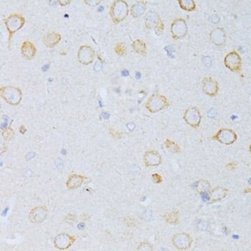
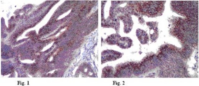
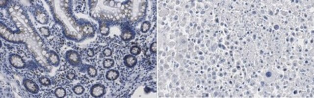
Immunohistochemistry Analysis: A representative lot detected CD63 (LAMP3) in Immunohistochemistry applications (Li, J., et. al. (2003). J Immunol. 171(6):2922-9).
Research Category
Inflammation & Immunology

Qualità

Evaluated by Western Blotting in human platelet lysate.

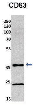
Western Blotting Analysis: 2 µg/mL of this antibody detected CD63 (LAMP3) in human platelet lysate.

Descrizione del bersaglio

~43 kDa observed; 25.64 kDa calculated. Uncharacterized bands may be observed in some lysate(s).

Stato fisico

Format: Purified
Protein G purified
Purified mouse monoclonal antibody IgG1 in buffer containing 0.1 M Tris-Glycine (pH 7.4), 150 mM NaCl with 0.05% sodium azide.

Stoccaggio e stabilità

Stable for 1 year at 2-8°C from date of receipt.

Altre note

Concentration: Please refer to lot specific datasheet.

Esclusione di responsabilità

Unless otherwise stated in our catalog or other company documentation accompanying the product(s), our products are intended for research use only and are not to be used for any other purpose, which includes but is not limited to, unauthorized commercial uses, in vitro diagnostic uses, ex vivo or in vivo therapeutic uses or any type of consumption or application to humans or animals.

Non trovi il prodotto giusto?  

Prova il nostro Motore di ricerca dei prodotti.


Certificati d'analisi (COA)

Cerca il Certificati d'analisi (COA) digitando il numero di lotto/batch corrispondente. I numeri di lotto o di batch sono stampati sull'etichetta dei prodotti dopo la parola ‘Lotto’ o ‘Batch’.

Possiedi già questo prodotto?

I documenti relativi ai prodotti acquistati recentemente sono disponibili nell’Archivio dei documenti.

Visita l’Archivio dei documenti

Il team dei nostri ricercatori vanta grande esperienza in tutte le aree della ricerca quali Life Science, scienza dei materiali, sintesi chimica, cromatografia, discipline analitiche, ecc..

Contatta l'Assistenza Tecnica.