
AB5407
Anti-Opsin Antibody, blue
Chemicon®, from rabbit
Sinonimo/i:
Anti-BCP, Anti-BOP
About This Item
Prodotti consigliati


Origine biologica
rabbit
Livello qualitativo
Forma dell’anticorpo
purified immunoglobulin
Tipo di anticorpo
primary antibodies
Clone
polyclonal
Reattività contro le specie
monkey, human, mouse
Produttore/marchio commerciale
Chemicon®
tecniche
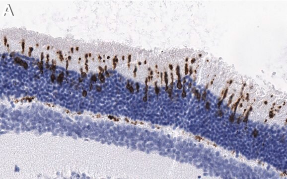
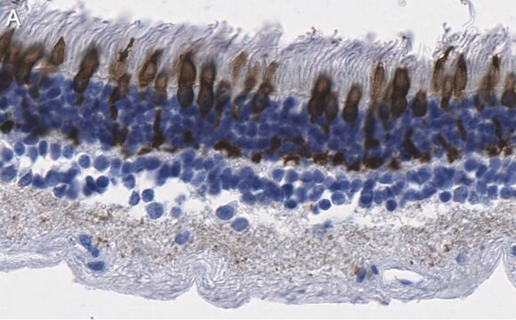
immunohistochemistry: suitable (paraffin)
N° accesso NCBI
N° accesso UniProt
Condizioni di spedizione
wet ice
modifica post-traduzionali bersaglio
unmodified
Informazioni sul gene
human ... OPN1LW(5956)
Descrizione generale
Specificità
Immunogeno
Applicazioni
Optimal working dilutions must be determined by the end user.
Neuroscience
Sensory & PNS
Stato fisico
Stoccaggio e stabilità
Risultati analitici
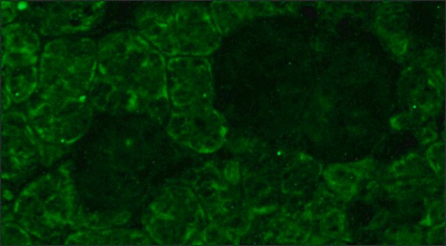
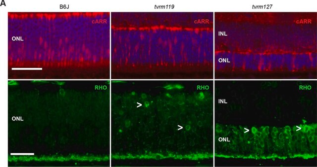
Retina
Note legali
Esclusione di responsabilità
Non trovi il prodotto giusto?
Prova il nostro Motore di ricerca dei prodotti.
Codice della classe di stoccaggio
12 - Non Combustible Liquids
Classe di pericolosità dell'acqua (WGK)
WGK 1
Punto d’infiammabilità (°F)
Not applicable
Punto d’infiammabilità (°C)
Not applicable
Certificati d'analisi (COA)
Cerca il Certificati d'analisi (COA) digitando il numero di lotto/batch corrispondente. I numeri di lotto o di batch sono stampati sull'etichetta dei prodotti dopo la parola ‘Lotto’ o ‘Batch’.
Possiedi già questo prodotto?
I documenti relativi ai prodotti acquistati recentemente sono disponibili nell’Archivio dei documenti.
Il team dei nostri ricercatori vanta grande esperienza in tutte le aree della ricerca quali Life Science, scienza dei materiali, sintesi chimica, cromatografia, discipline analitiche, ecc..
Contatta l'Assistenza Tecnica.