Skip to Content
Merck
All Photos(1)

Key Documents

C7706

Sigma-Aldrich

Monoclonal Anti-VSV Glycoprotein−Cy3 antibody produced in mouse

clone P5D4, purified immunoglobulin, buffered aqueous solution

Synonym(s):

Monoclonal Anti-VSV Glycoprotein

Sign Into View Organizational & Contract Pricing


About This Item

UNSPSC Code:
12352203
NACRES:
NA.46

biological source

mouse

Quality Level

conjugate

CY3 conjugate

antibody form

purified immunoglobulin

antibody product type

primary antibodies

clone

P5D4, monoclonal

form

buffered aqueous solution

technique(s)

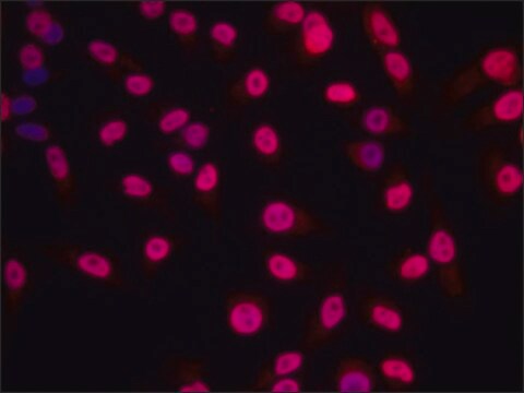
direct immunofluorescence: 1:10,000 using COS-7 cells transfected with a VSV-G tagged vinculin construct

isotype

IgG1

shipped in

wet ice

storage temp.

2-8°C

target post-translational modification

unmodified

Looking for similar products? Visit Product Comparison Guide

General description

Monoclonal Anti-VSV Glycoprotein (mouse IgG1 isotype) is derived from the P5D4 hybridoma produced by the fusion of mouse myeloma cells and splenocytes from a BALB/c mouse immunized with a synthetic peptide of vesicular stomatitis virus glycoprotein (VSV-G), conjugated to keyhole limpet hemocyanin (KLH). VSV-G is a protein expressed on the surface of bullet shaped virions.

Specificity

The antibody recognizes an epitope containing the five carboxy-terminal amino acids of VSV Glycoprotein. In infected cells, the antibody localizes the immature forms of VSV-G in the rough endoplasmic reticulum (RER) and in the cisternae of Golgi complex, as well as mature VSV-G at the cell surface and in the budding virus. The antibody does not stain the secreted form of VSV-G which lacks the membrane and the cytoplasmic domain. This antibody has been used for studies on the role of the cytoplasmic domain on newly-synthesized VSV-G during transfer to the plasma membrane and cell surface, using micro-injected antibody, immunoblotting, immunoprecipitation, immunocytochemistry and immunoelectron microscopy. The antibody has been used for the detection, immunoprecipitation and immunocytochemical staining of exogenously introduced constructs tagged with the carboxyl-terminus of VSV-G. This tag does not interfere with the function of the studied protein and can be specifically recognized by the P5D4 antibody without cross-reaction with any endogenous protein.

Immunogen

synthetic peptide containing the 15 carboxy-terminal amino acids (497-511) of Vesicular Stomatitis Virus Glycoprotein (VSV-G), conjugated to KLH.

Application

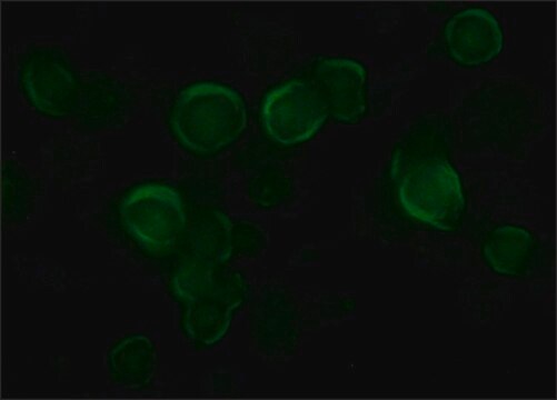
A dilution of 1:1000 of Monoclonal Anti-VSV Glycoprotein-Cy3® antibody produced in mouse was used to detect VSV glycoprotein in pig sperms by immunofluorescence.
Monoclonal Anti-VSV Glycoprotein Cy3 antibody produced in mouse has been used:
  • in studies applying
  • microinjection of antibody
  • immunoblotting
  • immunoprecipitation
  • immunocytochemistry
  • immunoelectron microscopy.

Biochem/physiol Actions

The envelope of vesicular stomatitis virus (VSV) consists of a bilayer membrane with a single type of glycoprotein, the G-protein (VSV-G) which mediates attachment to the cell surface and induces pH-dependent fusion between viral and target membranes. The carboxyl terminus of the VSV-G protein which does not have any homology with cellular proteins, has been engineered into expression vectors as a tag. Proteins expressed with this tag may thus be detected and localized using an antibody reactive specifically against this epitope with no risk of cellular background staining.
The vesicular stomatitis virus (VSV) glycoprotein facilitates the entry of HIV into the CD4 T cells through endocytotic pathway. The coupling of Cy3 to Anti-VSV Glycoprotein antibody allows for the visualization of protein by fluorescent microscopy.

Physical form

Solution in 0.01 M phosphate buffered saline, pH 7.4, containing 15 mM sodium azide

Legal Information

Cy is distributed under license from Amersham Biosciences Limited.
Cy is a registered trademark of Cytiva
Cy3 is a trademark of Cytiva

Disclaimer

Unless otherwise stated in our catalog or other company documentation accompanying the product(s), our products are intended for research use only and are not to be used for any other purpose, which includes but is not limited to, unauthorized commercial uses, in vitro diagnostic uses, ex vivo or in vivo therapeutic uses or any type of consumption or application to humans or animals.

Not finding the right product?  

Try our Product Selector Tool.

Storage Class Code

10 - Combustible liquids

WGK

nwg

Flash Point(F)

Not applicable

Flash Point(C)

Not applicable

Personal Protective Equipment

dust mask type N95 (US), Eyeshields, Gloves

Certificates of Analysis (COA)

Search for Certificates of Analysis (COA) by entering the products Lot/Batch Number. Lot and Batch Numbers can be found on a product’s label following the words ‘Lot’ or ‘Batch’.

Already Own This Product?

Find documentation for the products that you have recently purchased in the Document Library.

Visit the Document Library

Long-term, targeted genetic modification of the aqueous humor outflow tract coupled with noninvasive imaging of gene expression in vivo
Loewen N, et al.
Investigative Ophthalmology & Visual Science, 45(9), 3091-3098 (2004)
Nils Loewen et al.
Investigative ophthalmology & visual science, 45(9), 3091-3098 (2004-08-25)
To address a problem impeding research into glaucoma-associated genetic mutations and glaucoma gene therapy and achieve permanent, targeted transgene expression in the trabecular meshwork (TM). Lentiviral vectors are known to transduce human donor eye TM ex vivo, but efficacy in
Satoshi Aoki et al.
Nephrology, dialysis, transplantation : official publication of the European Dialysis and Transplant Association - European Renal Association, 33(1), 26-33 (2017-10-11)
Heparan sulphate proteoglycan (HSPG) is present in the glomerular basement membrane (GBM) and is thought to play a major role in the glomerular charge barrier. Reductions and structural alterations of HSPG are observed in different types of kidney diseases accompanied
Laura Miesen et al.
Disease models & mechanisms, 15(3) (2021-12-21)
In the glomerulus, Bowman's space is formed by a continuum of glomerular epithelial cells. In focal segmental glomerulosclerosis (FSGS), glomeruli show segmental scarring, a result of activated parietal epithelial cells (PECs) invading the glomerular tuft. The segmental scars interrupt the
Mechanism of membrane fusion induced by vesicular stomatitis virus G protein
Kim IS, et al.
Proceedings of the National Academy of Sciences of the USA, 114(1), E28-E36 (2017)

Our team of scientists has experience in all areas of research including Life Science, Material Science, Chemical Synthesis, Chromatography, Analytical and many others.

Contact Technical Service