Skip to Content
Merck
All Photos(2)

Key Documents

C0618

Sigma-Aldrich

Anti-Calsequestrin-1 antibody produced in rabbit

enhanced validation

~1.6 mg/mL, affinity isolated antibody, buffered aqueous solution

Synonym(s):

Anti-Aspartactin, Anti-CALMITINE, Anti-CASQ1, Anti-Calsequestrin-skeletal muscle isoform

Sign Into View Organizational & Contract Pricing


About This Item

MDL number:
UNSPSC Code:
12352203
NACRES:
NA.41

biological source

rabbit

Quality Level

conjugate

unconjugated

antibody form

affinity isolated antibody

antibody product type

primary antibodies

clone

polyclonal

form

buffered aqueous solution

mol wt

antigen ~60 kDa

species reactivity

canine, mouse, human, rat

enhanced validation

recombinant expression
Learn more about Antibody Enhanced Validation

concentration

~1.6 mg/mL

technique(s)

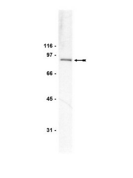
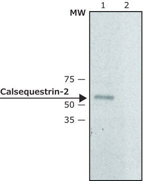
western blot: 0.5-1 μg/mL using extracts of rat skeletal muscle S1 fraction

UniProt accession no.

shipped in

dry ice

storage temp.

−20°C

target post-translational modification

unmodified

Gene Information

human ... CASQ1(844)
mouse ... Casq1(12372)

General description

Calsequestrin is expected to modulate ryanodine receptor (RyR) signaling and Ca2+ release with the help of triadin and junction proteins.

Immunogen

synthetic peptide corresponding to amino acids 66-84 located near the N-terminus of mouse calsequestrin-1, conjugated to KLH. This sequence is identical in human, rat, and dog calsequestrin-1 and highly conserved in bovine (single amino acid substitution). This sequence is not found in calsequestrin-2.

Application

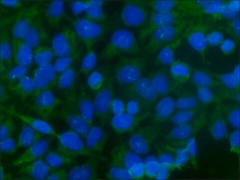
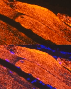
Anti-Calsequestrin-1 antibody produced in rabbit has been used in immunofluorescence.
Anti-Calsequestrin-1 antibody produced in rabbit is suitable for western blotting at a concentration of 0.5-1μg/mL using extracts of rat skeletal muscle S1 fraction.

Biochem/physiol Actions

Calsequestrin-1 is a putative auto-antigen associated with eye muscle inflammation in Graves′ disease. Calsequestrin-2 (55 kDa) is found in the SR′s terminal cisternae luminal space of cardiac, slow skeletal muscle cells and may be in platelets. The major Ca2+ binding protein in cardiac and skeletal muscle is a high-capacity, low-affinity Ca2+ binding glycoprotein, which functions as an internal Ca2+ store in the lumen of the SR. It helps in controlling the smooth-muscle contraction. Calsequestrin is expected to modulate ryanodine receptor (RyR) signaling and Ca2+ release with the help of triadin and junction proteins.†

Target description

Calsequestrin (CS also known as CSQ), the major Ca2+ binding protein in cardiac and skeletal muscle, is a high-capacity, low-affinity Ca2+ binding glycoprotein, which function as an internal Ca2+ store in the lumen of the SR.1-3 In mammals, two forms of the protein exist, calsequestrin-1 (CASQ-1) and calsequestrin-2 (CASQ-2), which encode the fast-twitch skeletal muscle and cardiac calsequestrin, respectively. Calsequestrin-1 is found in the SRs terminal cisternae luminal space of fast skeletal muscle cells. Calsequestrin-1 is a putative autoantigen associated with eye muscle inflammation in Graves′ disease.

Physical form

Solution in 0.01 M phosphate buffered saline, pH 7.4, containing 15 mM sodium azide.

Disclaimer

Unless otherwise stated in our catalog or other company documentation accompanying the product(s), our products are intended for research use only and are not to be used for any other purpose, which includes but is not limited to, unauthorized commercial uses, in vitro diagnostic uses, ex vivo or in vivo therapeutic uses or any type of consumption or application to humans or animals.

Not finding the right product?  

Try our Product Selector Tool.

Storage Class Code

10 - Combustible liquids

WGK

WGK 3

Flash Point(F)

Not applicable

Flash Point(C)

Not applicable

Personal Protective Equipment

dust mask type N95 (US), Eyeshields, Gloves

Certificates of Analysis (COA)

Search for Certificates of Analysis (COA) by entering the products Lot/Batch Number. Lot and Batch Numbers can be found on a product’s label following the words ‘Lot’ or ‘Batch’.

Already Own This Product?

Find documentation for the products that you have recently purchased in the Document Library.

Visit the Document Library

Identification of cellular pathogenicity markers for SIL1 mutations linked to Marinesco-Sjogren syndrome
Gatz C, et al.
Frontiers in Neurology, 10 (2019)
K Gunji et al.
Autoimmunity, 29(1), 1-9 (1999-03-03)
It is generally accepted that thyroid-associated ophthalmopathy (TAO) is an autoimmune disease of the eye muscle (EM) and the surrounding orbital connective tissue in which circulating antibodies play an important role. Antibodies against EM membrane proteins of 63-67kDa mol. wt.
Calsequestrin, triadin and more: the molecules that modulate calcium release in cardiac and skeletal muscle
Rios E and Gyorke S
The Journal of Physiology, 587(Pt 13), 3069-3069 (2009)
B Gopinath et al.
Clinical and experimental immunology, 145(1), 56-62 (2006-06-24)
We have identified several eye muscle antigens and studied the significance of the corresponding serum autoantibodies in patients with Graves' disease. Of these antigens, only calsequestrin is expressed more in eye muscle than other skeletal muscles, which could explain at
A O Jorgensen et al.
The Journal of cell biology, 98(4), 1597-1602 (1984-04-01)
Localization of calsequestrin in chicken ventricular muscle cells was determined by indirect immunofluorescence and immuno-Protein A-colloidal gold labeling of cryostat and ultracryotomy sections, respectively. Calsequestrin was localized in the lumen of peripheral junctional sarcoplasmic reticulum, as well as in the

Our team of scientists has experience in all areas of research including Life Science, Material Science, Chemical Synthesis, Chromatography, Analytical and many others.

Contact Technical Service