General description
Lipopolysaccharide-responsive and beige-like anchor protein (UniProt: P50851; also known as Beige-like protein, CDC4-like protein) is encoded by the LRBA (also known as BGL, CDC4L, LBA) gene (Gene ID: 987) in human. LRBA is a single-pass membrane protein that displays ubiquitous expression in normal tissues, but its expression is elevated in malignancy. In immune cells. It is found in all membrane compartments that are associated with receptor signaling, including the plasma membrane, Golgi complex, clathrin-coated pits, and trans-endocytic vesicles. Its expression is induced in B cells and macrophages by bacterial lipopolysaccharides (LPS). Two isoforms of LRBA have been reported that are produced by alternative splicing. LRBA contains 2 BEACH (Beige and Chediak-Higashi) domains and six WD repeats at its C-terminal. LRBA may be involved in coupling signal transduction and vesicle trafficking to enable polarized secretion and/or membrane deposition of immune effector molecules. It coordinates signaling of immune receptors to promote effector function and plays a crucial role in immune regulation. Mutations in LRBA gene are associated with defective B-cell differentiation and decreased or absent antibody production. (Ref.: Park, MY et al. (2016). Nat. Scientific Reports 6:36568).
Specificity
Clone 2D4.1 detects human Lipopolysaccharide-responsive and beige-like anchor protein (LRBA). It targets an epitope within 115 amino acids from the N-terminal half.
Immunogen
GST/His-tagged recombinant fragment corresponding to 115 amino acids from the N-terminal half of human Lipopolysaccharide-responsive and beige-like anchor protein (LRBA).
Application
Anti-LRBA, clone 2D4.1 Antibody, Cat. No. MABF1096 is a mouse monoclonal antibody that detects Lipopolysaccharide-responsive and beige-like anchor protein (LRBA) and has been tested for use in Immunohistochemistry (Paraffin) and Western Blotting.
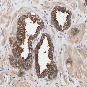
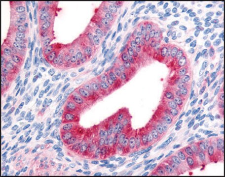
Immunohistochemistry Analysis: A 1:250 dilution from a representative lot detected LRBA in human pancreas, human testis, and human cervical cancer tissue sections.
Research Category
Inflammation & Immunology
Quality
Evaluated by Western Blotting in Jurkat cell membrane preparations.
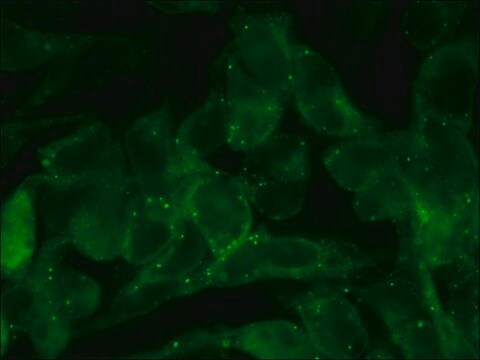
Western Blotting Analysis: 1 µg/mL of this antibody detected LRBA in 10 µg of Jurkat cell membrane preparations.
Target description
~320 kDa observed; 319.11 kDa calculated. Uncharacterized bands may be observed in some lysate(s).
Physical form
Format: Purified
Protein G purified
Purified mouse monoclonal antibody IgG1 in buffer containing 0.1 M Tris-Glycine (pH 7.4), 150 mM NaCl with 0.05% sodium azide.
Storage and Stability
Stable for 1 year at 2-8°C from date of receipt.
Other Notes
Concentration: Please refer to lot specific datasheet.
Disclaimer
Unless otherwise stated in our catalog or other company documentation accompanying the product(s), our products are intended for research use only and are not to be used for any other purpose, which includes but is not limited to, unauthorized commercial uses, in vitro diagnostic uses, ex vivo or in vivo therapeutic uses or any type of consumption or application to humans or animals.