Skip to Content
Merck
All Photos(3)

Documents

ABN26

Sigma-Aldrich

Anti-iNOS/NOS II Antibody, NT

from rabbit, purified by affinity chromatography

Synonym(s):

Nitric oxide synthase, inducible, Inducible NO synthase, Inducible NOS, iNOS, Macrophage NOS, MAC-NOS, NOS type II, Peptidyl-cysteine S-nitrosylase NOS2

Sign Into View Organizational & Contract Pricing


About This Item

UNSPSC Code:
12352203
eCl@ss:
32160702
NACRES:
NA.41

biological source

rabbit

Quality Level

antibody form

affinity isolated antibody

antibody product type

primary antibodies

clone

polyclonal

purified by

affinity chromatography

species reactivity

mouse, human

packaging

antibody small pack of 25 μg

technique(s)

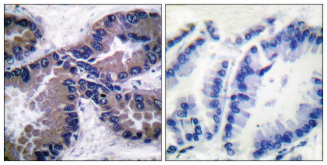
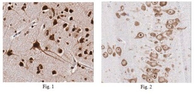
immunohistochemistry: suitable (paraffin)
immunoprecipitation (IP): suitable
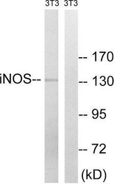
western blot: suitable

NCBI accession no.

UniProt accession no.

shipped in

ambient

storage temp.

2-8°C

target post-translational modification

unmodified

Gene Information

mouse ... Nos2(18126)

General description

Nitric oxide (NO) is an inorganic, gaseous free radical that carries a variety of messages between cells. Vasorelaxation, neurotransmission and cytotoxicity can all be potentiated through cellular response to NO. NO production is mediated by members of the nitric oxide synthase (NOS) family. NOS catalyzes the oxidization of L-arginine to produce L-citrulline and NO. Two constitutive isoforms, brain or neuronal NOS (b or nNOS, type I) & endothelial cell NOS (eNOS, type III), and one inducible isoform (iNOS, type II), have been cloned. Cytokines such as interferon-gamma (IFN), tumor necrosis factor (TNF), interleukin-1 and -2, and lipopolysaccarides (LPS) cause an increase in iNOS mRNA, protein, and activity levels. Protein kinase C-stimulating agents exhibit the same effect on iNOS activity. Human iNOS is regulated by calcium/calmodulin (in contrast with mouse NOS2).

Specificity

Other homologies: Rat (85% sequence homology).
This antibody recognizes iNOS/NOS II at the N-terminus.

Immunogen

Epitope: N-terminus
GST-tagged recombinant protein corresponding to the N-terminus of mouse iNOS/NOS II.

Application

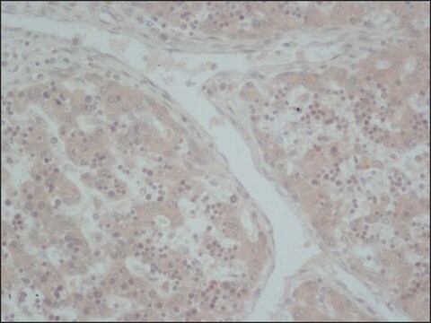
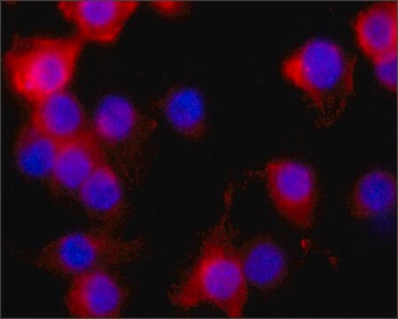
Detect iNOS/NOS II using this Anti-iNOS/NOS II Antibody, NT validated for use in WB, IP, IH(P).
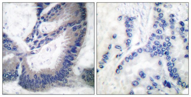
Immunohistochemistry Analysis: 1:50-200 dilution from a representative lot detected iNOS/NOS II in malignant human lung tissues.

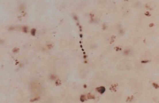
Immunoprecipitation Analysis: 10 µg of this antibody immunoprecipitated iNOS/NOSII from IFNgamma/LPS treated RAW264.7 cell lysate.
Research Category
Neuroscience
Research Sub Category
Oxidative Stress

Quality

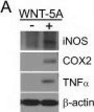
Evaluated by Western Blot in IFNgamma/LPS untreated and treated RAW264.7 cell lysates.

Western Blot Analysis: 0.5 µg/mL of this antibody detected iNOS/NOS II on 10 µg of IFNgamma/LPS untreated and treated RAW264.7 cell lysates.

Target description

~125 kDa observed

Linkage

Replaces: 06-573

Physical form

Affinity purified
Purified rabbit polyclonal in buffer containing 0.1 M Tris-Glycine (pH 7.4), 150 mM NaCl with 0.05% sodium azide.

Storage and Stability

Stable for 1 year at 2-8°C from date of receipt.

Analysis Note

Control
IFNgamma/LPS untreated and treated RAW264.7 cell lysates

Other Notes

Concentration: Please refer to the Certificate of Analysis for the lot-specific concentration.

Disclaimer

Unless otherwise stated in our catalog or other company documentation accompanying the product(s), our products are intended for research use only and are not to be used for any other purpose, which includes but is not limited to, unauthorized commercial uses, in vitro diagnostic uses, ex vivo or in vivo therapeutic uses or any type of consumption or application to humans or animals.

Not finding the right product?  

Try our Product Selector Tool.


Certificates of Analysis (COA)

Search for Certificates of Analysis (COA) by entering the products Lot/Batch Number. Lot and Batch Numbers can be found on a product’s label following the words ‘Lot’ or ‘Batch’.

Already Own This Product?

Find documentation for the products that you have recently purchased in the Document Library.

Visit the Document Library

Francisco J Salguero et al.
Parasites & vectors, 11(1), 73-73 (2018-02-02)
Visceral leishmaniasis (VL) is a neglected tropical disease (NTD), caused by the intracellular protozoan parasites Leishmania donovani and Leishmania infantum. Symptomatic VL is considered fatal when left untreated. At present, there is no effective vaccine licensed for human use and
Florence Vallelian et al.
Pharmacology research & perspectives, 6(2), e00392-e00392 (2018-04-04)
Activation of the innate immune system by free heme has been proposed as one of the principal consequences of cell-free hemoglobin (Hb) exposure. Nonetheless, in the absence of infection, heme exposures within a hematoma, during hemolysis, or upon systemic administration
Delphine Séhédic et al.
Theranostics, 7(18), 4517-4536 (2017-11-22)
Gold standard beam radiation for glioblastoma (GBM) treatment is challenged by resistance phenomena occurring in cellular populations well prepared to survive or to repair damage caused by radiation. Among signals that have been linked with radio-resistance, the SDF1/CXCR4 axis, associated
Kosaku Okuda et al.
Nature communications, 14(1), 621-621 (2023-02-05)
DNA methyltransferases (DNMTs) catalyze methylation at the C5 position of cytosine with S-adenosyl-L-methionine. Methylation regulates gene expression, serving a variety of physiological and pathophysiological roles. The chemical mechanisms regulating DNMT enzymatic activity, however, are not fully elucidated. Here, we show
Gulsan Ara Sathi et al.
Journal of cell science, 130(9), 1559-1569 (2017-03-30)
The importance of macrophages in tissue development and regeneration has been strongly emphasized. However, the specific roles of macrophage colony-stimulating factor (MCSF), the key regulator of macrophage differentiation, in glandular tissue development have been unexplored. Here, we disclose new macrophage-independent

Our team of scientists has experience in all areas of research including Life Science, Material Science, Chemical Synthesis, Chromatography, Analytical and many others.

Contact Technical Service