Skip to Content
Merck
All Photos(2)

Documents

AV35098

Sigma-Aldrich

Anti-KCNN4 antibody produced in rabbit

affinity isolated antibody

Synonym(s):

Anti-IK1, Anti-IKCA1, Anti-KCA4, Anti-KCa3.1, Anti-SK4, Anti-Small conductance calcium-activated channel, subfamily N, member 4, Anti-hIKCa1, Anti-hKCa4

Sign Into View Organizational & Contract Pricing


About This Item

UNSPSC Code:
12352203
NACRES:
NA.41

biological source

rabbit

Quality Level

conjugate

unconjugated

antibody form

affinity isolated antibody

antibody product type

primary antibodies

clone

polyclonal

mol wt

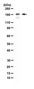
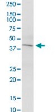
48 kDa

species reactivity

human

concentration

0.5 mg - 1 mg/mL

technique(s)

western blot: suitable

NCBI accession no.

UniProt accession no.

shipped in

wet ice

storage temp.

−20°C

target post-translational modification

unmodified

Gene Information

human ... KCNN4(3783)

General description

Potassium intermediate/small conductance calcium-activated channel, subfamily N, member 4, KCNN4, is a component of a potassium channel activated by calcium. It is critically involved in calcium ion transport and cell homeostasis.

Immunogen

Synthetic peptide directed towards the C terminal region of human KCNN4

Application

Anti-KCNN4 antibody produced in rabbit has been used in:
  • western blot analysis
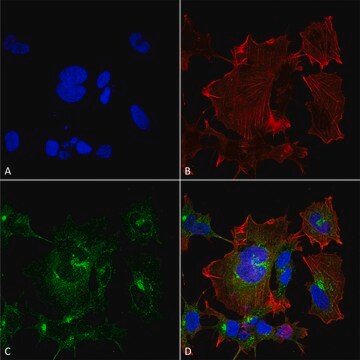
  • immunofluorescence
  • staining
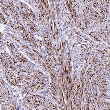
  • immunohistochemistry

Biochem/physiol Actions

KCNN4 (potassium calcium-activated channel subfamily N member 4) is part of a potentially heterotetrameric voltage-independent potassium channel that is activated by intracellular calcium. Activation is followed by membrane hyperpolarization, which promotes calcium influx. KCNN4 may be part of the predominant calcium-activated potassium channel in T-lymphocytes. Activation is followed by membrane hyperpolarization, which promotes calcium influx. The encoded protein may be part of the predominant calcium-activated potassium channel in T-lymphocytes. This gene is similar to other KCNN family potassium channel genes, but it differs enough to possibly be considered as part of a new subfamily.

Sequence

Synthetic peptide located within the following region: DLQQNLSSSHRALEKQIDTLAGKLDALTELLSTALGPRQLPEPSQQSK

Physical form

Purified antibody supplied in 1x PBS buffer with 0.09% (w/v) sodium azide and 2% sucrose.

Disclaimer

Unless otherwise stated in our catalog or other company documentation accompanying the product(s), our products are intended for research use only and are not to be used for any other purpose, which includes but is not limited to, unauthorized commercial uses, in vitro diagnostic uses, ex vivo or in vivo therapeutic uses or any type of consumption or application to humans or animals.

Not finding the right product?  

Try our Product Selector Tool.

Storage Class Code

10 - Combustible liquids

WGK

WGK 2

Flash Point(F)

Not applicable

Flash Point(C)

Not applicable


Certificates of Analysis (COA)

Search for Certificates of Analysis (COA) by entering the products Lot/Batch Number. Lot and Batch Numbers can be found on a product’s label following the words ‘Lot’ or ‘Batch’.

Already Own This Product?

Find documentation for the products that you have recently purchased in the Document Library.

Visit the Document Library

Epigenetic dysregulation of KCa3. 1 channels induces poor prognosis in lung cancer
Bulk E, et al.
International Journal of Cancer. Journal International Du Cancer, 137(6), 1306-1317 (2015)
A Novel Gene, hKCa4, Encodes the Calcium-activated Potassium Channel in Human T Lymphocytes
Logsdon NJ, et al.
The Journal of Biological Chemistry, 272(52), 32723-32726 (1997)
Bradykinin-induced chemotaxis of human gliomas requires the activation of KCa3. 1 and ClC-3
Cuddapah VA, et al.
The Journal of Neuroscience, 33(4), 1427-1440 (2013)
D Strøbæk et al.
British journal of pharmacology, 168(2), 432-444 (2012-08-16)
The K(Ca) 3.1 channel is a potential target for therapy of immune disease. We identified a compound from a new chemical class of K(Ca) 3.1 inhibitors and assessed in vitro and in vivo inhibition of immune responses. We characterized the
Yi-Je Chen et al.
Journal of cerebral blood flow and metabolism : official journal of the International Society of Cerebral Blood Flow and Metabolism, 36(12), 2146-2161 (2015-12-15)
Activated microglia/macrophages significantly contribute to the secondary inflammatory damage in ischemic stroke. Cultured neonatal microglia express the K

Our team of scientists has experience in all areas of research including Life Science, Material Science, Chemical Synthesis, Chromatography, Analytical and many others.

Contact Technical Service