EF5012
Anti-EF5 Antibody, clone ELK3-51 Antibody, Cyanine 5 conjugate
clone ELK3-51, from mouse, CY5 conjugate
About This Item
Recommended Products
biological source
mouse
conjugate
CY5 conjugate
antibody product type
primary antibodies
clone
ELK3-51, monoclonal
species reactivity
human, rat, mouse
technique(s)
immunofluorescence: suitable
immunohistochemistry: suitable
isotype
IgG1
General description
Advantages of the EF5 hypoxia detection method:
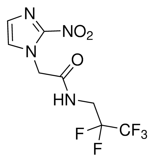
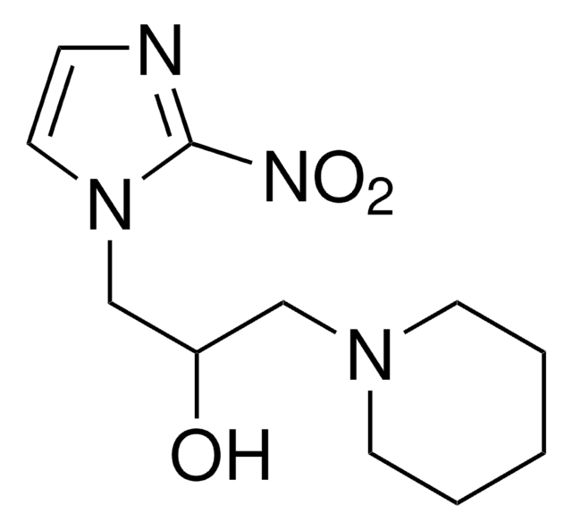
• EF5 exists in only one form; Pimidozole, an alternative hypoxia marker, exists in two forms; one of which is charged and very hydrophilic, the other lipophilic. Pimidozole thus has a very complex biodistribution. In contrast, EF5 is lipophilic and uncharged and this allows very rapid and even tissue distribution.
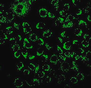
• EF5 binding images can be calibrated to provide quantitative data on the pO2 values of each cell (1). The fluorescent images obtained from EF5 binding can be calibrated according to camera settings and a “cube-binding” value which is obtained through a separate procedure. The intensity values of calibrated images are directly related to actual tissue pO2 values. As a result, these images provide information regarding not only where hypoxic areas may or may not be, but also data regarding the distribution and levels of hypoxia.
Reference:
Koch CJ (2002) Measurement of absolute oxygen levels in cells and tissues using oxygen sensors and 2-nitroimidazole EF5. Methods in Enzymology 352: 3-31.
Application
Cancer
Hypoxia
Storage and Stability
Other Notes
Disclaimer
Not finding the right product?
Try our Product Selector Tool.
Storage Class Code
12 - Non Combustible Liquids
WGK
WGK 2
Flash Point(F)
Not applicable
Flash Point(C)
Not applicable
Certificates of Analysis (COA)
Search for Certificates of Analysis (COA) by entering the products Lot/Batch Number. Lot and Batch Numbers can be found on a product’s label following the words ‘Lot’ or ‘Batch’.
Already Own This Product?
Find documentation for the products that you have recently purchased in the Document Library.
Articles
Hypoxia detection assays to measure oxygen levels in both live and fixed cells and tissues.
Our team of scientists has experience in all areas of research including Life Science, Material Science, Chemical Synthesis, Chromatography, Analytical and many others.
Contact Technical Service