Skip to Content
Merck
All Photos(3)

Key Documents

HPA016938

Sigma-Aldrich

Anti-ATP6V0D1 antibody produced in rabbit

enhanced validation

Prestige Antibodies® Powered by Atlas Antibodies, affinity isolated antibody, buffered aqueous glycerol solution

Synonym(s):

Anti-32 kDa accessory protein, Anti-P39, Anti-V-ATPase 40 kDa accessory protein, Anti-V-ATPase AC39 subunit, Anti-V-ATPase subunit d 1, Anti-Vacuolar proton pump subunit d 1

Sign Into View Organizational & Contract Pricing


About This Item

UNSPSC Code:
12352203
Human Protein Atlas Number:
NACRES:
NA.41

biological source

rabbit

conjugate

unconjugated

antibody form

affinity isolated antibody

antibody product type

primary antibodies

clone

polyclonal

product line

Prestige Antibodies® Powered by Atlas Antibodies

form

buffered aqueous glycerol solution

species reactivity

human

enhanced validation

recombinant expression
Learn more about Antibody Enhanced Validation

technique(s)

immunoblotting: 0.04-0.4 μg/mL
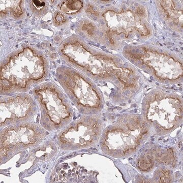
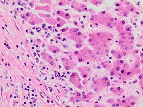
immunohistochemistry: 1:20-1:50

immunogen sequence

TELSKEDRAKLFPHCGRLYPEGLAQLARADDYEQVKNVADYYPEYKLLFEGAGSNPGDKTLEDRFFEHEVKLNKLAFLNQFHFGVFYAFVKL

UniProt accession no.

shipped in

wet ice

storage temp.

−20°C

target post-translational modification

unmodified

Gene Information

human ... ATP6V0D1(9114)

Immunogen

Vacuolar proton pump subunit d1(V-ATPase subunit d 1) (V-ATPase AC39 subunit) (V-ATPase 40 kDa accessory protein) (P39) (32 kDa accessory protein).

Application

Applications in which this antibody has been used successfully, and the associated peer-reviewed papers, are given below.
Western Blotting (1 paper)

Biochem/physiol Actions

ATP6V0D1 (ATPase, H+ transporting, lysosomal 38kDa, V0 subunit d1) is a subunit D of the vacuolar proton ATPase. It is expressed in kidney and placenta. ATP6V0D1 mediates the migration of protons into intracellular organelles or across the plasma membrane of specialized cells such as osteoclast and renal intercalated cells. V-ATPase combined with phosphoinositide-binding proteins, regulate vesicular trafficking pathway during ciliogenesis.

Features and Benefits

Prestige Antibodies® are highly characterized and extensively validated antibodies with the added benefit of all available characterization data for each target being accessible via the Human Protein Atlas portal linked just below the product name at the top of this page. The uniqueness and low cross-reactivity of the Prestige Antibodies® to other proteins are due to a thorough selection of antigen regions, affinity purification, and stringent selection. Prestige antigen controls are available for every corresponding Prestige Antibody and can be found in the linkage section.

Every Prestige Antibody is tested in the following ways:
  • IHC tissue array of 44 normal human tissues and 20 of the most common cancer type tissues.
  • Protein array of 364 human recombinant protein fragments.

Linkage

Corresponding Antigen APREST71568

Physical form

Solution in phosphate-buffered saline, pH 7.2, containing 40% glycerol and 0.02% sodium azide

Legal Information

Prestige Antibodies is a registered trademark of Merck KGaA, Darmstadt, Germany

Disclaimer

Unless otherwise stated in our catalog or other company documentation accompanying the product(s), our products are intended for research use only and are not to be used for any other purpose, which includes but is not limited to, unauthorized commercial uses, in vitro diagnostic uses, ex vivo or in vivo therapeutic uses or any type of consumption or application to humans or animals.

Not finding the right product?  

Try our Product Selector Tool.

Storage Class Code

10 - Combustible liquids

WGK

WGK 1

Flash Point(F)

Not applicable

Flash Point(C)

Not applicable

Personal Protective Equipment

dust mask type N95 (US), Eyeshields, Gloves

Certificates of Analysis (COA)

Search for Certificates of Analysis (COA) by entering the products Lot/Batch Number. Lot and Batch Numbers can be found on a product’s label following the words ‘Lot’ or ‘Batch’.

Already Own This Product?

Find documentation for the products that you have recently purchased in the Document Library.

Visit the Document Library

A K Agarwal et al.
Biochemical and biophysical research communications, 279(2), 543-547 (2000-12-19)
The HSD11B2 and VPATPD genes encoding the human kidney isozyme of 11beta-hydroxysteroid dehydrogenase (11-HSD2) and subunit D of the vacuolar proton ATPase, respectively, are located on chromosome 16q22. They are transcribed from complementary DNA strands and their 3' ends are
Zhang Bao et al.
International journal of molecular medicine, 38(3), 823-833 (2016-07-20)
Mycobacterium tuberculosis (MTB), one of the major bacterial pathogens for lethal infectious diseases, is capable of surviving within the phagosomes of host alveolar macrophages; therefore, host genetic variations may alter the susceptibility to MTB. In this study, to identify host
Yanqun Chen et al.
Cell research, 22(2), 333-345 (2011-08-17)
Sorting nexins (SNXs) are phosphoinositide-binding proteins implicated in the sorting of various membrane proteins in vitro, but the in vivo functions of them remain largely unknown. We reported previously that SNX10 is a unique member of the SNX family genes
B van Hille et al.
Biochemical and biophysical research communications, 197(1), 15-21 (1993-11-30)
The vacuolar proton ATPase (V-ATPase) translocates protons into intracellular organelles or across the plasma membrane of specialised cells such as osteoclast and renal intercalated cells. The catalytic site of the V-ATPase consists of a hexamer of three A subunits and

Our team of scientists has experience in all areas of research including Life Science, Material Science, Chemical Synthesis, Chromatography, Analytical and many others.

Contact Technical Service