Skip to Content
Merck
All Photos(1)

Documents

AV30475

Sigma-Aldrich

Anti-BCL2L1 antibody produced in rabbit

IgG fraction of antiserum

Synonym(s):

Anti-BCL-XL/S, Anti-BCL2-like 1, Anti-BCL2L, Anti-BCLX, Anti-Bcl-X, Anti-Bcl-xL, Anti-Bcl-xS, Anti-DKFZp781P2092

Sign Into View Organizational & Contract Pricing


About This Item

MDL number:
UNSPSC Code:
12352203
NACRES:
NA.41

biological source

rabbit

Quality Level

conjugate

unconjugated

antibody form

IgG fraction of antiserum

antibody product type

primary antibodies

clone

polyclonal

form

buffered aqueous solution

mol wt

26 kDa

species reactivity

sheep, rabbit, horse, pig, human, bovine, dog

concentration

0.5 mg - 1 mg/mL

technique(s)

western blot: suitable

NCBI accession no.

UniProt accession no.

shipped in

wet ice

storage temp.

−20°C

target post-translational modification

unmodified

Gene Information

human ... BCL2L1(598)

General description

BCL2L1 is a signaling molecule that is known to regulate neuronal apoptosis. Furthermore, BCL2L1 can facilitate the survival of retinal ganglion cells.
Rabbit Anti-BCL2L1 associates with rabbit, rat, bovine, canine, pig, mouse, and human BCL2L1.

Immunogen

Synthetic peptide directed towards the N terminal region of human BCL2L1

Application

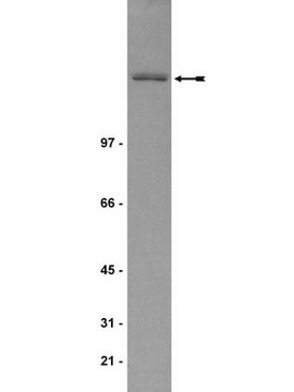
Rabbit Anti-BCL2L1 antibody can be used for western blot (2.5μg/ml) assays.

Biochem/physiol Actions

BCL2L1 encodes a protein which belongs to the BCL-2 protein family. The proteins encoded by BCL2L1 are located at the outer mitochondrial membrane, and have been shown to regulate outer mitochondrial membrane channel (VDAC) opening. VDAC regulates mitochondrial membrane potential, and thus controls the production of reactive oxygen species and release of cytochrome C by mitochondria, both of which are the potent inducers of cell apoptosis.The protein encoded by this gene belongs to the BCL-2 protein family. BCL-2 family members form hetero- or homodimers and act as anti- or pro-apoptotic regulators that are involved in a wide variety of cellular activities. The proteins encoded by this gene are located at the outer mitochondrial membrane, and have been shown to regulate outer mitochondrial membrane channel (VDAC) opening. VDAC regulates mitochondrial membrane potential, and thus controls the production of reactive oxygen species and release of cytochrome C by mitochondria, both of which are the potent inducers of cell apoptosis. Two alternatively spliced transcript variants, which encode distinct isoforms, have been reported. The longer isoform acts as an apoptotic inhibitor and the shorter form acts as an apoptotic activator.

Sequence

Synthetic peptide located within the following region: SNRELVVDFLSYKLSQKGYSWSQFSDVEENRTEAPEGTESEMETPSAING

Physical form

Purified antibody supplied in 1x PBS buffer with 0.09% (w/v) sodium azide and 2% sucrose.

Disclaimer

Unless otherwise stated in our catalog or other company documentation accompanying the product(s), our products are intended for research use only and are not to be used for any other purpose, which includes but is not limited to, unauthorized commercial uses, in vitro diagnostic uses, ex vivo or in vivo therapeutic uses or any type of consumption or application to humans or animals.

Not finding the right product?  

Try our Product Selector Tool.

Storage Class Code

10 - Combustible liquids

WGK

WGK 3

Flash Point(F)

Not applicable

Flash Point(C)

Not applicable


Certificates of Analysis (COA)

Search for Certificates of Analysis (COA) by entering the products Lot/Batch Number. Lot and Batch Numbers can be found on a product’s label following the words ‘Lot’ or ‘Batch’.

Already Own This Product?

Find documentation for the products that you have recently purchased in the Document Library.

Visit the Document Library

Joanna Janisiak et al.
International journal of molecular sciences, 22(15) (2021-08-08)
Rhabdomyosarcoma (RMS) is a malignant soft tissue cancer that develops mostly in children and young adults. With regard to histopathology, four rhabdomyosarcoma types are distinguishable: embryonal, alveolar, pleomorphic and spindle/sclerosing. Currently, increased amounts of evidence indicate that not only gene
Jeffrey M Harder et al.
Molecular and cellular neurosciences, 51(1-2), 53-59 (2012-07-28)
The Bcl-2 family is responsible for regulating cell death pathways in neurons during development, after injury and in disease. The activation of the pro-death family member BAX is often the final step before cell death in neurons. Pro-survival family members
Li Gao et al.
Hematology (Amsterdam, Netherlands), 19(3), 148-157 (2013-07-31)
Bone marrow stromal cells (BMSCs) can support multiple myeloma (MM) disease progression and resistance to chemotherapy. The proliferation of MM cells may be suppressed by modifying the hematopoietic microenvironment (HME). We have previously isolated human umbilical cord blood-derived stromal cells
W He et al.
Oncogene, 33(23), 3004-3013 (2013-07-09)
Killing cancer cells through the induction of apoptosis is one of the main mechanisms of chemotherapy. However, numerous cancer cells have primary or acquired apoptosis resistance, resulting in chemoresistance. In this study, using a novel chalcone derivative chalcone-24 (Chal-24), we
Yong Xin et al.
Molecular medicine reports, 10(3), 1569-1575 (2014-07-26)
High‑dose total body irradiation (TBI) has an established role as preparative regimen for bone‑marrow transplantation in the treatment of chronic myelogenous leukemia (CML), but this regimen still has a relatively high rate of acute and late toxicity. Low‑dose radiation (LDR)

Our team of scientists has experience in all areas of research including Life Science, Material Science, Chemical Synthesis, Chromatography, Analytical and many others.

Contact Technical Service