Skip to Content
Merck
All Photos(1)

Key Documents

328R-1

Sigma-Aldrich

PU.1 (EPR3158Y) Rabbit Monoclonal Primary Antibody

Sign Into View Organizational & Contract Pricing


About This Item

UNSPSC Code:
12352203
NACRES:
NA.41

biological source

rabbit

Quality Level

100
500

conjugate

unconjugated

antibody form

culture supernatant

antibody product type

primary antibodies

clone

EPR3158Y, monoclonal

description

For In Vitro Diagnostic Use in Select Regions (See Chart)

form

buffered aqueous solution

species reactivity

human

packaging

vial of 0.1 mL concentrate (328R-14)
vial of 0.5 mL concentrate (328R-15)
bottle of 1.0 mL predilute (328R-17)
vial of 1.0 mL concentrate (328R-16)
bottle of 7.0 mL predilute (328R-18)

manufacturer/tradename

Cell Marque

technique(s)

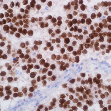
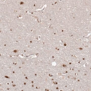
immunohistochemistry (formalin-fixed, paraffin-embedded sections): 1:50-1:200

isotype

IgG

control

tonsil

shipped in

wet ice

storage temp.

2-8°C

visualization

nuclear

Gene Information

human ... SPI1(6688)

General description

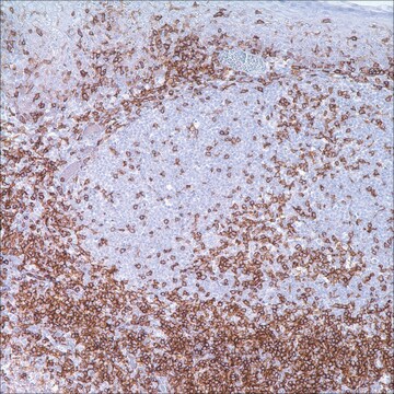
PU.1 is a transcription factor that has been shown to be important for normal B-cell development. PU.1 belongs to the ETS family of transcription factors. It is expressed in the myeloid lineage and in immature as well as mature B-lymphocytes, with the exception of plasma cells. PU.1 is essential during early B-cell differentiation. The absence of PU.1 results in total block of B-cell development at the pre-pro stage. Very little is known about PU.1 function in later stages of B-cell development. PU.1 does not seem to play a role in the end-stage of B-cell development and is not expressed in plasma cells. PU.1 exerts an important role in the regulation of the expression of crucial B-cell proteins, such as immunoglobulin (Ig) genes, and CD20 and its putative binding sites were also identified in the promoters of CD79, CD10, and CD22. PU.1 binds to the 3′ enhancer region of both the Ig kappa and lambda light chain genes and it also regulates the immunoglobulin heavy chain genes through the intron enhancer region. PU.1 is expressed in germinal center B-cells and mantle B-cells. Various lymphomas are also positive for this marker including the following: B-chronic lymphocytic leukemia, mantle cell lymphoma, follicular lymphoma, marginal zone lymphoma, Burkitt lymphoma, diffuse large cell lymphoma, diffuse large B-cell lymphoma, T-cell rich B-cell lymphoma, and nodular lymphocyte predominant Hodgkin lymphoma.

Quality


IVD

IVD

IVD

RUO

Linkage

PU.1 Positive Control Slides, Product No. 328S, are available for immunohistochemistry (formalin-fixed, paraffin-embedded sections).

Physical form

Solution in Tris Buffer, pH 7.3-7.7, with 1% BSA and <0.1% Sodium Azide

Preparation Note

Download the IFU specific to your product lot and formatNote: This requires a keycode which can be found on your packaging or product label.

Other Notes

For Technical Service please contact: 800-665-7284 or email: service@cellmarque.com

Legal Information

Cell Marque is a trademark of Merck KGaA, Darmstadt, Germany

Not finding the right product?  

Try our Product Selector Tool.


Choose from one of the most recent versions:

Certificates of Analysis (COA)

Lot/Batch Number

Don't see the Right Version?

If you require a particular version, you can look up a specific certificate by the Lot or Batch number.

Already Own This Product?

Find documentation for the products that you have recently purchased in the Document Library.

Visit the Document Library

Christoph Loddenkemper et al.
The Journal of pathology, 202(1), 60-69 (2003-12-25)
It has previously been demonstrated that in cultured and in situ tumour cells of classical Hodgkin lymphoma (cHL), the immunoglobulin (Ig) promoter is inactive and its transcription factors Oct2 and/or BOB.1/OBF.1 are down-regulated. In this study, the analysis of these
R Hromas et al.
Blood, 82(10), 2998-3004 (1993-11-15)
The ETS oncogene family member PU.1 is a transcriptional activator that is dysregulated by Friend erythroleukemia virus insertion. Northern analysis found that PU.1 is highly expressed in cells of myeloid and B-lymphoid origin, but not expressed at all in a
Juliette J Hoefnagel et al.
Modern pathology : an official journal of the United States and Canadian Academy of Pathology, Inc, 19(9), 1270-1276 (2006-06-17)
Expression patterns of eight transcription factors involved in different stages of B-cell development were investigated in a large group of primary cutaneous B-cell lymphomas and compared with expression patterns during normal B-cell development. The following transcription factors were investigated: Pax-5

Our team of scientists has experience in all areas of research including Life Science, Material Science, Chemical Synthesis, Chromatography, Analytical and many others.

Contact Technical Service