Skip to Content
Merck
All Photos(1)

Key Documents

MABN323

Sigma-Aldrich

Anti-Ermin Antibody, clone 160

clone 160, from mouse

Synonym(s):

Ermin, Juxtanodin

Sign Into View Organizational & Contract Pricing


About This Item

UNSPSC Code:
12352203
eCl@ss:
32160702
NACRES:
NA.41

biological source

mouse

Quality Level

antibody form

purified immunoglobulin

antibody product type

primary antibodies

clone

160, monoclonal

species reactivity

mouse, rat

technique(s)

immunocytochemistry: suitable
immunohistochemistry: suitable
western blot: suitable

isotype

IgG2aκ

NCBI accession no.

UniProt accession no.

shipped in

wet ice

target post-translational modification

unmodified

Gene Information

mouse ... Ermn(77767)

Related Categories

General description

Ermin is also known as Juxtanodin (JN). Ermin is involved in cytoskeletal rearrangements during myelinogenesis and in the maintenance and stability of the myelin sheath in adults. Ermin is also thought to be important in oligodendroglia maturation, myelin/Ranvier node formation during CNS development, and in the plasticity of related structures in the mature CNS. Ermin is exclusively expressed by oligodendrocytes in the brain and spinal cord, where it appears in the late stages of myelination, and in the mature nerves, it is localized in the outer cytoplasmic lip of the myelin sheath and the paranodal loops.

Immunogen

Recombinant protein corresponding to mouse Ermin.

Application

Anti-Ermin Antibody, clone 160 is a highly specific mouse monoclonal antibody, that targets Ermin & has been tested in western blotting, ICC & IHC.
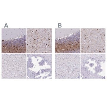
Immunohistochemistry Analysis: A representative lot from an independent laboratory detected Ermin in mouse wild type sagittal brain sections, and demonstrated a loss of signal in mouse OL-ablated sagittal brain sections (Brockschnieder, D., et al. (2006). J Neurosci. 26(3):737-762).

Immunocytochemistry Analysis: A representative lot from an independent laboratory detected Ermin in certain mouse brain and optic nerve sections (Brockschnieder, D., et al. (2006). J Neurosci. 26(3):737-762).
Research Category
Neuroscience
Research Sub Category
Developmental Neuroscience

Quality

Evaluated by Western Blotting in mouse brain tissue lysate.

Western Blotting Analysis: 1 µg/mL of this antibody detected Ermin in 10 µg of mouse brain tissue lysate.

Target description

~42 kDa observed. The calculated molecular weight of this protein is 32 kDa; however, it has been observed at ~42 kDa (Brockschnieder, D., et al. (2006). J Neurosci. 26(3):737-76.).

Physical form

Format: Purified
Protein G Purified
Purified mouse monoclonal IgG2aκ in buffer containing 0.1 M Tris-Glycine (pH 7.4), 150 mM NaCl with 0.05% sodium azide.

Storage and Stability

Stable for 1 year at 2-8°C from date of receipt.

Analysis Note

Control
Mouse brain tissue lysate

Other Notes

Concentration: Please refer to the Certificate of Analysis for the lot-specific concentration.

Disclaimer

Unless otherwise stated in our catalog or other company documentation accompanying the product(s), our products are intended for research use only and are not to be used for any other purpose, which includes but is not limited to, unauthorized commercial uses, in vitro diagnostic uses, ex vivo or in vivo therapeutic uses or any type of consumption or application to humans or animals.

Not finding the right product?  

Try our Product Selector Tool.

Storage Class Code

12 - Non Combustible Liquids

WGK

WGK 1

Flash Point(F)

Not applicable

Flash Point(C)

Not applicable


Certificates of Analysis (COA)

Search for Certificates of Analysis (COA) by entering the products Lot/Batch Number. Lot and Batch Numbers can be found on a product’s label following the words ‘Lot’ or ‘Batch’.

Already Own This Product?

Find documentation for the products that you have recently purchased in the Document Library.

Visit the Document Library

Damian Brockschnieder et al.
The Journal of neuroscience : the official journal of the Society for Neuroscience, 26(3), 757-762 (2006-01-20)
Oligodendrocytes form an insulating multilamellar structure of compact myelin around axons, thereby allowing rapid propagation of action potentials. Despite the considerable clinical importance of myelination, little is known about the molecular mechanisms that enable oligodendrocytes to generate their specialized membrane
Carola I Radulescu et al.
Neurobiology of disease, 135, 104744-104744 (2020-01-14)
Structural and molecular myelination deficits represent early pathological features of Huntington disease (HD). Recent evidence from germ-free (GF) animals suggests a role for microbiota-gut-brain bidirectional communication in the regulation of myelination. In this study, we aimed to investigate the impact

Our team of scientists has experience in all areas of research including Life Science, Material Science, Chemical Synthesis, Chromatography, Analytical and many others.

Contact Technical Service