MAB4211
Anti-CD34 Class I Antibody, clone B1-3C5
clone B1-3C5, Chemicon®, from mouse
Sign Into View Organizational & Contract Pricing
All Photos(1)
About This Item
UNSPSC Code:
12352203
eCl@ss:
32160702
NACRES:
NA.41
Recommended Products
biological source
mouse
Quality Level
antibody form
purified antibody
antibody product type
primary antibodies
clone
B1-3C5, monoclonal
species reactivity
human
manufacturer/tradename
Chemicon®
technique(s)
flow cytometry: suitable
immunofluorescence: suitable
isotype
IgG1
NCBI accession no.
UniProt accession no.
shipped in
wet ice
target post-translational modification
unmodified
Gene Information
human ... CD34(947)
Related Categories
Specificity
This antibody belongs to CD34 (assigned by the Third International Workshop on Leukocyte Differentiation Antigens, Oxford, 1987; Hogg & Horton, 1987). It reacts with a 120kD glycoprotein found on progenitor cell populations in human bone marrow and foetal liver (Katz et al., 1985). This antigen is normally restricted to immature human haemopoietic precursor cells (Peschel & Koller, 1989).
Cell reactivity (Tindle et al., 1985):
Normal: No reaction with normal peripheral lymphocytes, granulocytes, monocytes, erythrocytes and platelets. Reacts with <4% of cells in normal bone marrow.
Clinical: Reaction
Acute myeloid leukemia (AML, M1 and M2) positive
Null lymphoblastic leukemia (Null ALL) positive
Common acute lymphoblastic leukemia (CALL) positive
Myeloblastic and lymphoblastic crises of chronic
granulocytic leukemia (CGL) positive
Chronic myeloid leukemia (CML) negative
T cell acute lymphoblastic leukemia (TALL) negative
B cell acute lymphoblastic leukemia (BALL) negative
Chronic lymphocytic leukemia (CLL) negative
Cell lines:
Reacts with early myeloblast line KG1. No reaction with null, B or T lymphoid, erythroid or later myeloid cell lines.
Immunogen
KG1 human acute myelogenous cell line.
Application
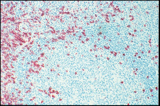
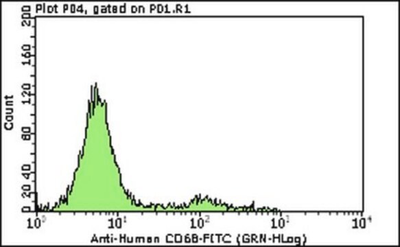
Detect CD34 Class I using this Anti-CD34 Antibody (Class 1), clone B1-3C5 validated for use in FC & IF.
Identification of CD34 positive cells is important for the diagnosis and classification of acute myeloid leukemia (AML). It is especially useful in patients with poorly differentiated myeloblasts, as well as in myeloblastic crises of chronic granulocytic leukemia. It has been recommended that this antibody be used as part of a panel for screening of leukemia (Batinic et al., 1989).
SUGGESTED USAGE DILUTION
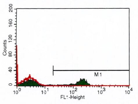
1. Flow cytometry and indirect immunofluorescence 1:25
Dilute with isotonic buffer. Use 50 μl of diluted antibody per 1 x 10E6 peripheral blood mononuclear cells (PBMC) or bone marrow cells in 100 μl of buffer.
2. Indirect immunoperoxidase staining - the final dilution will depend on the assay conditions and detection system employed. However, a dilution of at least 1:25 will be applicable to most commonly used systems.
Research Category
Cell Structure
Cell Structure
Research Sub Category
ECM Proteins
ECM Proteins
Physical form
Format: Purified
The antibody is supplied in phosphate buffered saline, pH 7.4, containing 0.2% bovine serum albumin and 0.1% sodium azide. The characteristics of each lot are tested by electrophoresis and flow cytometry.
Storage and Stability
For prolonged periods, store below -20°C in undiluted aliquots. AVOID REPEATED FREEZE/THAW CYCLES. Product stable for 6 months when stored as directed.
WARNING: The monoclonal reagent solution contains 0.1% sodium azide as a preservative. Due to potential hazards arising from the build up of this material in pipes, spent reagent should be disposed of with liberal volumes of water.
WARNING: The monoclonal reagent solution contains 0.1% sodium azide as a preservative. Due to potential hazards arising from the build up of this material in pipes, spent reagent should be disposed of with liberal volumes of water.
Legal Information
CHEMICON is a registered trademark of Merck KGaA, Darmstadt, Germany
Disclaimer
Unless otherwise stated in our catalog or other company documentation accompanying the product(s), our products are intended for research use only and are not to be used for any other purpose, which includes but is not limited to, unauthorized commercial uses, in vitro diagnostic uses, ex vivo or in vivo therapeutic uses or any type of consumption or application to humans or animals.
Not finding the right product?
Try our Product Selector Tool.
Storage Class Code
12 - Non Combustible Liquids
WGK
WGK 2
Certificates of Analysis (COA)
Search for Certificates of Analysis (COA) by entering the products Lot/Batch Number. Lot and Batch Numbers can be found on a product’s label following the words ‘Lot’ or ‘Batch’.
Already Own This Product?
Find documentation for the products that you have recently purchased in the Document Library.
A phase I clinical trial of the treatment of Crohn's fistula by adipose mesenchymal stem cell transplantation.
Garcia-Olmo, Damian, et al.
Diseases of the Colon and Rectum, 48, 1416-1423 (2005)
In-Su Park et al.
PloS one, 10(6), e0122776-e0122776 (2015-06-13)
We investigated whether low-level light irradiation prior to transplantation of adipose-derived stromal cell (ASC) spheroids in an animal skin wound model stimulated angiogenesis and tissue regeneration to improve functional recovery of skin tissue. The spheroid, composed of hASCs, was irradiated
Our team of scientists has experience in all areas of research including Life Science, Material Science, Chemical Synthesis, Chromatography, Analytical and many others.
Contact Technical Service