Skip to Content
Merck
All Photos(2)

Key Documents

07-1719

Sigma-Aldrich

Anti-FOXO3A Antibody

from rabbit, purified by affinity chromatography

Synonym(s):

AF6q21 protein, Forkhead in rhabdomyosarcoma-like 1, forkhead box O3, forkhead box O3A, forkhead homolog (rhabdomyosarcoma) like 1

Sign Into View Organizational & Contract Pricing


About This Item

UNSPSC Code:
12352203
eCl@ss:
32160702
NACRES:
NA.41
clone:
polyclonal
application:
ICC
WB
species reactivity:
human
technique(s):
immunocytochemistry: suitable
western blot: suitable
citations:
8

biological source

rabbit

Quality Level

antibody form

affinity isolated antibody

antibody product type

primary antibodies

clone

polyclonal

purified by

affinity chromatography

species reactivity

human

species reactivity (predicted by homology)

mouse

technique(s)

immunocytochemistry: suitable
western blot: suitable

NCBI accession no.

UniProt accession no.

shipped in

wet ice

target post-translational modification

unmodified

Gene Information

human ... FOXO3(2309)

General description

Forkhead box O3A (FOXO3A) is a member of the forkhead family of transcription factors (FOXO), which play an important role in cellular proliferation, metabolism, stress tolerance, and aging. FOXO transcription factors have a characteristic forkhead domain and are located in the cytoplasm. Upon oxidative stress, FOXO3A translocates to the nucleus where binding to the DNA sequence 5’-[AG]TAAA[TC]A-3’ occurs and IGF-1 is dephosphlorated, resulting in apoptosis. In order for the cell to survive, IGF-1 must be phosphlorated by AKT1/PKB at Thr32 and Ser-253, which will result in retention and inactivation of FOXO3A in the cytoplasm. Translocation of FOXO3A with the MLL gene has been associated with secondary acute leukemia.

Specificity

This antibody recognizes FOXO3A.

Immunogen

Synthetic peptide corresponding to FOX03A

Application

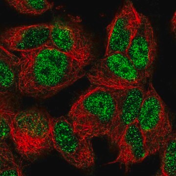
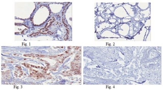
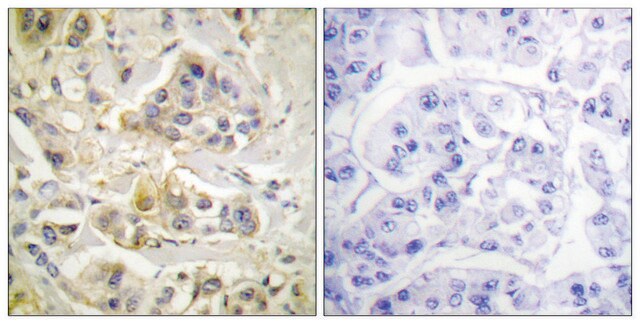
Immunocytochemistry:
A 1:500 dilution of this antibody was used to detect FOXO3A in HeLa cells. Positive staining in the nucleus.
Research Category
Signaling
Research Sub Category
Insulin/Energy Signaling

Transcription Factors
This Anti-FOXO3A Antibody is validated for use in WB, IC for the detection of FOXO3A.

Quality

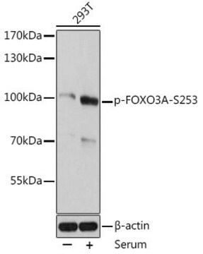
Western Blot Analysis:
1:500 dilution of this antibody was used to detect FOXO3A in Jurkat cell lysate.

Target description

~80 kDa

Linkage

Replaces: 04-1007

Physical form

Affinity purified
Purified rabbit polyclonal antibody precipitated in a solution of 50% saturated ammonium sulfate and PBS containing no preservatives.

Storage and Stability

Stable for 1 year at 2-8ºC from date of receipt.
To reconstitute the antibody, centrifuge the antibody at a moderate speed (5,000 rpm) for 5 minutes. Carefully remove the ammonium sulfate/PBS buffer solution and discard; 10μL of residual ammonium sulfate solution will not affect the re-suspension of the antibody. Do not let the protein pellet dry, as severe loss of antibody reactivity can occur. Re-suspend the antibody pellet in 100L either standard PBS or TBS (pH 7.3-7.5). DO NOT VORTEX. Mix by gentle stirring with a wide pipet tip or gentle finger-tapping. Let the precipitated antibody rehydrate for 1 hour at 25°C prior to use. Small particles of precipitated antibody that fail to re-suspend are normal. Vials are overfilled to compensate for any losses.

Analysis Note

Control
FOXO3A in Jurkat cell lysate.

Disclaimer

Unless otherwise stated in our catalog or other company documentation accompanying the product(s), our products are intended for research use only and are not to be used for any other purpose, which includes but is not limited to, unauthorized commercial uses, in vitro diagnostic uses, ex vivo or in vivo therapeutic uses or any type of consumption or application to humans or animals.

Not finding the right product?  

Try our Product Selector Tool.

Storage Class Code

12 - Non Combustible Liquids

WGK

WGK 2

Flash Point(F)

Not applicable

Flash Point(C)

Not applicable


Regulatory Listings

Regulatory Listings are mainly provided for chemical products. Only limited information can be provided here for non-chemical products. No entry means none of the components are listed. It is the user’s obligation to ensure the safe and legal use of the product.

EU REACH Annex XVII (Restriction List)

CAS No.

Certificates of Analysis (COA)

Search for Certificates of Analysis (COA) by entering the products Lot/Batch Number. Lot and Batch Numbers can be found on a product’s label following the words ‘Lot’ or ‘Batch’.

Already Own This Product?

Find documentation for the products that you have recently purchased in the Document Library.

Visit the Document Library

FOXO3A directs a protective autophagy program in haematopoietic stem cells.
Warr, MR; Binnewies, M; Flach, J; Reynaud, D; Garg, T; Malhotra, R; Debnath, J; Passegue, E
Nature null
The E-box binding factors Max/Mnt, MITF, and USF1 act coordinately with FoxO to regulate expression of proapoptotic and cell cycle control genes by phosphatidylinositol 3-kinase/Akt/glycogen synthase kinase 3 signaling.
Terragni, J; Nayak, G; Banerjee, S; Medrano, JL; Graham, JR; Brennan, JF; Sepulveda, S; Cooper, GM
The Journal of Biological Chemistry null
Stephen E McGowan et al.
Respiratory research, 14, 68-68 (2013-07-04)
Secondary pulmonary alveolar septal formation requires platelet derived growth factor (PDGF-A) and platelet derived growth factor receptor-alpha (PDGFRα), and their regulation influences alveolar septal areal density and thickness. Insufficient PDGFRα expression in lung fibroblasts (LF) results in failed septation. Mice
Roy Kalfon et al.
Cardiovascular research, 113(2), 134-146 (2017-01-14)
Obesity and type 2 diabetes (T2D) trigger a harmful stress-induced cardiac remodeling process known as cardiomyopathy. These diseases represent a serious and widespread health problem in the Western world; however the underlying molecular basis is not clear. ATF3 is an

Our team of scientists has experience in all areas of research including Life Science, Material Science, Chemical Synthesis, Chromatography, Analytical and many others.

Contact Technical Service