Skip to Content
Merck
All Photos(8)

Documents

HPA022040

Sigma-Aldrich

Anti-RUNX2 antibody produced in rabbit

enhanced validation

Prestige Antibodies® Powered by Atlas Antibodies, affinity isolated antibody, buffered aqueous glycerol solution

Synonym(s):

Anti-Acute myeloid leukemia 3 protein, Anti-CBF-alpha-1, Anti-Core-binding factor subunit alpha-1, Anti-OSF-2, Anti-Oncogene AML-3, Anti-PEA2-alpha A, Anti-PEBP2-alpha A, Anti-Polyomavirus enhancer-binding protein 2 alpha A subunit, Anti-Runt-related transcription factor 2, Anti-SL3-3 enhancer factor 1 alpha A subunit, Anti-SL3/AKV core-binding factor alpha A subunit

Sign Into View Organizational & Contract Pricing


About This Item

MDL number:
UNSPSC Code:
12352203
Human Protein Atlas Number:
NACRES:
NA.41

biological source

rabbit

conjugate

unconjugated

antibody form

affinity isolated antibody

antibody product type

primary antibodies

clone

polyclonal

product line

Prestige Antibodies® Powered by Atlas Antibodies

form

buffered aqueous glycerol solution

species reactivity

human

enhanced validation

RNAi knockdown
orthogonal RNAseq
Learn more about Antibody Enhanced Validation

technique(s)

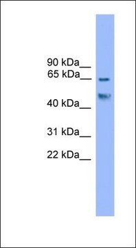
immunoblotting: 0.04-0.4 μg/mL
immunofluorescence: 0.25-2 μg/mL
immunohistochemistry: 1:200-1:500

immunogen sequence

LNSAPSPFNPQGQSQITDPRQAQSSPPWSYDQSYPSYLSQMTSPSIHSTTPLSSTRGTGLPAITDVPRRISGASELGPFSDPRQFPSISSLTESRFSNPRMHYPA

UniProt accession no.

application(s)

research pathology

shipped in

wet ice

storage temp.

−20°C

target post-translational modification

unmodified

Gene Information

human ... RUNX2(860)

General description

Runt-related transcription factor 2/acute myeloid leukemia 3 protein (RUNX2, AML-3) belongs to Runt DNA-binding domain transcription factor family. RUNX2 gene is mapped to human chromosome 6p21.1 RUNX2 possesses the Runt domain, nuclear localization signal (NLS), the nuclear matrix targeting signal (NMTS) and a C-terminal VWRPY sequence. It also has two additional domains, one being the N-terminal glutamine-alanine (QA) repeats and the other a C-terminal proline/serine/threonine (PST) rich tract. RUNX2 exists in two isoforms. The isoform 1 is expressed in osteoblasts and the isoform 2 is distributed in mesenchymal condensations and mature chondrocytes.
Runt-related transcription factor 2/acute myeloid leukemia 3 protein (RUNX2, AML-3) is a Runt DNA-binding domain transcription factor that facilitates osteoblast differentiation and skeletal morphogenesis.

Specificity

Rabbit polyclonal anti-RUNX2 antibody reacts with human runt-related transcription factor 2/Acute myeloid leukemia 3 protein.

Immunogen

Runt-related transcription factor 2 recombinant protein epitope signature tag (PrEST)

Application

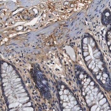
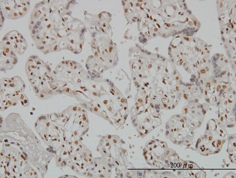
All Prestige Antibodies Powered by Atlas Antibodies are developed and validated by the Human Protein Atlas (HPA) project and as a result, are supported by the most extensive characterization in the industry.

The Human Protein Atlas project can be subdivided into three efforts: Human Tissue Atlas, Cancer Atlas, and Human Cell Atlas. The antibodies that have been generated in support of the Tissue and Cancer Atlas projects have been tested by immunohistochemistry against hundreds of normal and disease tissues and through the recent efforts of the Human Cell Atlas project, many have been characterized by immunofluorescence to map the human proteome not only at the tissue level but now at the subcellular level. These images and the collection of this vast data set can be viewed on the Human Protein Atlas (HPA) site by clicking on the Image Gallery link. We also provide Prestige Antibodies® protocols and other useful information.
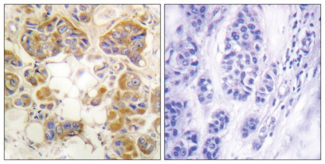
Anti-RUNX2 antibody produced in rabbit has been used in immunohistochemistry and immunofluorescence.
Rabbit polyclonal anti-RUNX2 antibody is used to tag Runt-related transcription factor 2/Acute myeloid leukemia 3 protein for detection and quantitation by immunocytochemical and immunohistochemical (IHC) techniques. It is used as a probe to determine the presence and roles of runt-related transcription factor 2/Acute myeloid leukemia 3 protein in osteoblast differentiation and skeletal morphogenesis.

Biochem/physiol Actions

Runt-related transcription factor 2/ acute myeloid leukemia 3 protein (RUNX2, AML-3) mediates osteoblast differentiation and skeletal morphogenesis. It promotes mesenchymal cell differentiation in osteoblasts and its maturation. Elevated levels of RUNX2 is observed in bone?metastatic cancers. The post-translational modifications like phosphorylation, methylation, glycosylation and ubiquitination in the RUNX2 modulates osteogenesis. Mutations in the RUNX2 gene leading to loss of function is implicated in cleidocranial dysplasia (CCD). The levels of RUNX2 is elevated in osteosarcoma.

Features and Benefits

Prestige Antibodies® are highly characterized and extensively validated antibodies with the added benefit of all available characterization data for each target being accessible via the Human Protein Atlas portal linked just below the product name at the top of this page. The uniqueness and low cross-reactivity of the Prestige Antibodies® to other proteins are due to a thorough selection of antigen regions, affinity purification, and stringent selection. Prestige antigen controls are available for every corresponding Prestige Antibody and can be found in the linkage section.

Every Prestige Antibody is tested in the following ways:
  • IHC tissue array of 44 normal human tissues and 20 of the most common cancer type tissues.
  • Protein array of 364 human recombinant protein fragments.

Linkage

Corresponding Antigen APREST86701

Physical form

Solution in phosphate-buffered saline, pH 7.2, containing 40% glycerol and 0.02% sodium azide

Legal Information

Prestige Antibodies is a registered trademark of Merck KGaA, Darmstadt, Germany

Disclaimer

Unless otherwise stated in our catalog or other company documentation accompanying the product(s), our products are intended for research use only and are not to be used for any other purpose, which includes but is not limited to, unauthorized commercial uses, in vitro diagnostic uses, ex vivo or in vivo therapeutic uses or any type of consumption or application to humans or animals.

Not finding the right product?  

Try our Product Selector Tool.

Storage Class Code

10 - Combustible liquids

WGK

WGK 1

Flash Point(F)

Not applicable

Flash Point(C)

Not applicable


Certificates of Analysis (COA)

Search for Certificates of Analysis (COA) by entering the products Lot/Batch Number. Lot and Batch Numbers can be found on a product’s label following the words ‘Lot’ or ‘Batch’.

Already Own This Product?

Find documentation for the products that you have recently purchased in the Document Library.

Visit the Document Library

Bianca Gazieri Castelucci et al.
PloS one, 13(4), e0195304-e0195304 (2018-04-06)
Animal models commonly serve as a bridge between in vitro experiments and clinical applications; however, few physiological processes in adult animals are sufficient to serve as proof-of-concept models for cartilage regeneration. Intriguingly, some rodents, such as young adult mice, undergo
Greg Holmes et al.
Gene expression patterns : GEP, 17(1), 16-25 (2014-12-17)
Sutures, where neighboring craniofacial bones are separated by undifferentiated mesenchyme, are major growth sites during craniofacial development. Pathologic fusion of bones within sutures occurs in a wide variety of craniosynostosis conditions and can result in dysmorphic craniofacial growth and secondary
Midface and upper airway dysgenesis in FGFR2-related craniosynostosis involves multiple tissue-specific and cell cycle effects
Holmes G, et al.
Development, 145(19), e0201492-e0201492 (2018)
Time-dependent regulation of morphological changes and cartilage differentiation markers in the mouse pubic symphysis during pregnancy and postpartum recovery
Castelucci BG, et al.
Testing, 13(4), e0195304-e0195304 (2018)
Neus Martínez-Abadías et al.
Disease models & mechanisms, 6(3), 768-779 (2013-03-23)
Apert syndrome is a congenital disorder characterized by severe skull malformations and caused by one of two missense mutations, S252W and P253R, on fibroblast growth factor receptor 2 (FGFR2). The molecular bases underlying differential Apert syndrome phenotypes are still poorly

Articles

Mesenchymal stem cell markers and antibodies suitable for investigating targets in fibroblasts, chondrocytes, adipocytes, osteoblasts, and muscle cells.

Our team of scientists has experience in all areas of research including Life Science, Material Science, Chemical Synthesis, Chromatography, Analytical and many others.

Contact Technical Service