Skip to Content
Merck
All Photos(6)

Key Documents

M1406

Sigma-Aldrich

Anti-MAP2 (2a+2b) antibody, Mouse monoclonal

clone AP-20, ascites fluid

Synonym(s):

Anti-Microtubule Associated Protein 2

Sign Into View Organizational & Contract Pricing


About This Item

MDL number:
UNSPSC Code:
12352203
NACRES:
NA.41

biological source

mouse

Quality Level

conjugate

unconjugated

antibody form

ascites fluid

antibody product type

primary antibodies

clone

AP-20, monoclonal

mol wt

apparent mol wt 280 kDa

contains

15 mM sodium azide

species reactivity

Xenopus, mouse, quail, human, bovine, rat, aquatic salamander

technique(s)

immunocytochemistry: suitable
western blot: 1:500 using rat brain enriched microtubule protein preparation or rat cerebral cortex extract

isotype

IgG1

UniProt accession no.

shipped in

dry ice

storage temp.

−20°C

target post-translational modification

unmodified

Gene Information

human ... MAP2(4133)
mouse ... Mtap2(17756)
rat ... Map2(25595)

Looking for similar products? Visit Product Comparison Guide

General description

Microtubule-associated protein 2 (MAP2) is a neuronal protein. It belongs to the MAP2/Tau family. This gene is located to human chromosome 2q34-q35. MAP2 is present in neurons. There are three forms of MAP2; two are similarly sized with apparent molecular weights of 280 kDa (MAP2a and MAP2b) and the third with a lower molecular weight of 70 kDa (MAP2c). In the newborn rat brain, MAP2b and MAP2c are present, while MAP2a is absent. Between postnatal days 10 and 20, MAP2a appears. At the same time, the level of MAP2c drops by 10-fold. This change happens during the period when dendrite growth is completed and when neurons have reached their mature morphology. MAP2 is degraded by a Cathepsin D-like protease in the brain of aged rats. There is some indication that MAP2 is expressed at higher levels in some types of neurons than in other types. MAP2 is known to promote microtubule assembly and to form side-arms on microtubules. It also interacts with neurofilaments, actin, and other elements of the cytoskeleton.
Monoclonal Anti-MAP2 (2a + 2b) (mouse IgG1 isotype) is derived from the hybridoma produced by the fusion of mouse myeloma cells and splenocytes from an immunized mouse.

Immunogen

bovine MAP2

Application

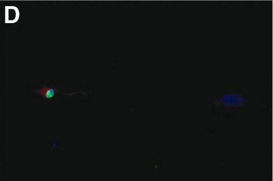
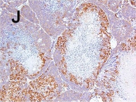
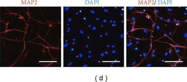
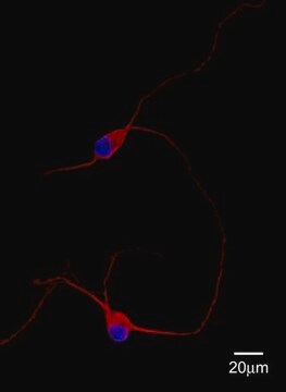
Monoclonal Anti-MAP2 (2a+2b) antibody has been used in immunohistochemistry, immunocytochemistry and antibody characterization.

Biochem/physiol Actions

Microtubule-associated protein 2 (MAP2) interacts with microtubules to maintain the structure of dendrites. It plays a major role in supporting the actin cytoskeleton in spines, binding and nucleating filamentous actin (f-actin) to modulate spine morphology.

Physical form

The product is provided as ascites fluid with 15 mM sodium azide as a preservative.

Storage and Stability

For continuous use, store at 2-8 °C for up to one month. For extended storage, the solution may be frozen in working aliquots. Repeated freezing and thawing is not recommended. Storage in "frost-free" freezers is not recommended. If slight turbidity occurs upon prolonged storage, clarify the solution by centrifugation before use.

Disclaimer

Unless otherwise stated in our catalog or other company documentation accompanying the product(s), our products are intended for research use only and are not to be used for any other purpose, which includes but is not limited to, unauthorized commercial uses, in vitro diagnostic uses, ex vivo or in vivo therapeutic uses or any type of consumption or application to humans or animals.

Not finding the right product?  

Try our Product Selector Tool.

Storage Class Code

11 - Combustible Solids

WGK

WGK 1


Certificates of Analysis (COA)

Search for Certificates of Analysis (COA) by entering the products Lot/Batch Number. Lot and Batch Numbers can be found on a product’s label following the words ‘Lot’ or ‘Batch’.

Already Own This Product?

Find documentation for the products that you have recently purchased in the Document Library.

Visit the Document Library

A Novel Purification Method for CNS Projection Neurons Leads to the Identification of Brain Vascular Cells as a Source of Trophic Support for Corticospinal Motor Neurons
Jason C D, et al.
The Journal of Neuroscience, 28(33) , 8294-8305 (2008)
The MAP2/Tau family of microtubule-associated proteins
Dehmelt L and Halpain S
Genome Biology, 6(1), 204-204 (2004)
Impaired Cerebellar Development and Function in Mice Lacking CAPS2, a Protein Involved in Neurotrophin Release
Tetsushi S, et al.
The Journal of Neuroscience, 27(10), 2472-2482 (2007)
Attenuated inflammatory response in triggering receptor expressed on myeloid cells 2 (TREM2) knock-out mice following stroke.
Sieber M W, et al.
PLoS ONE, 8(1), e52982-e52982 (2013)
Identification of ischemic regions in a rat model of stroke.
Popp A, et al.
PLoS ONE, 4(3), e4764-e4764 (2009)

Our team of scientists has experience in all areas of research including Life Science, Material Science, Chemical Synthesis, Chromatography, Analytical and many others.

Contact Technical Service