Skip to Content
Merck
All Photos(1)

Key Documents

B7653

Sigma-Aldrich

Monoclonal Anti-Biotin antibody produced in mouse

clone BN-34, ascites fluid

Synonym(s):

Monoclonal Anti-Biotin

Sign Into View Organizational & Contract Pricing


About This Item

UNSPSC Code:
12352203
NACRES:
NA.46

biological source

mouse

conjugate

unconjugated

antibody form

ascites fluid

antibody product type

primary antibodies

clone

BN-34, monoclonal

contains

15 mM sodium azide

technique(s)

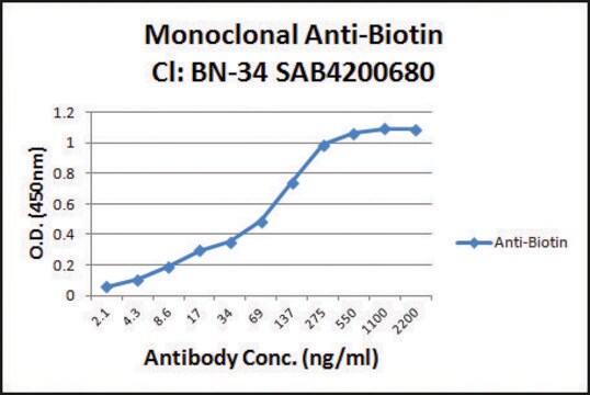
capture ELISA: suitable
immunohistochemistry (formalin-fixed, paraffin-embedded sections): suitable
indirect ELISA: 1:4,000

isotype

IgG1

application(s)

research pathology

shipped in

dry ice

storage temp.

−20°C

target post-translational modification

unmodified

Looking for similar products? Visit Product Comparison Guide

General description

Biotin is a water-soluble vitamin. It is also called as vitamin B8. Biotin is a bicyclic compound that has a uredo ring and a tetrahydrothiophene ring.
Monoclonal Anti-Biotin recognizes the free biotin molecule and biotin conjugated to various immunoglobulins in ELISA and immunohistochemical techniques. Specificity was verified by using biotinylated goat antibodies reactive against human and rabbit antigens coated on microwell plates.
The antibody recognizes free biotin and biocytin using competitive ELISA. Using indirect ELISA or immuno­histo­chemical staining, the antibody also recognizes biotin conjugated to various immunoglobulins. Specificity is evaluated using biotinylated goat antibodies to human and rabbit antigens coated on multiwell plates.

Immunogen

Biotin-KLH conjugate
Biotinylated keyhole limpet hemocyanin (KLH)

Application

Applications in which this antibody has been used successfully, and the associated peer-reviewed papers, are given below.
Western Blotting (1 paper)
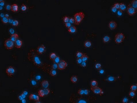
Monoclonal Anti-Biotin antibody has been used in proximity ligation assay (PLA), cell surface biotinylation assay and western blotting.
The antibody may be used in amplification techniques, ELISA, immunoblotting, in situ nucleic acid hybridization, flow cytometry, image analysis or confocal microscopy.
In some applications, localization of biotinylated probes with avidin produces high background levels. Anti-biotin reagents may be substituted for avidin to decrease non-specific binding.
Utilized in a variety of applications including blotting, immunocytochemistyr, insitu nucleic acid hybridization, ELISA, fluorescent activated cell-sorting (FACS) and electron microscopy.Titer is approximately 1:2000 by ELISA but should be optimized by the individual user for their application of choiceAn essential vitamin, biotin is required by living organisms and cells in culture. Avidin′s high affinity for biotin is often exploited in immuno-based assays. While standard assay methods using the avidin-biotin enzyme complex will suffice for most studies, there are occasions when enhanced sensitivity is needed to detect smaller amounts of antigen or localize low density antigens in histologic sections. Conventional immunoassay methods are improved by the use of Monoclonal Anti-Biotin which enhances the sensitivity of avidin-biotin immunoassays by bridging a second layer of avidin-biotin-enzyme complex. This antibody can be used in many other applications where biotin can be introduced as a target label.
Monoclonal Anti-Biotin antibody produced in mouse is suitable for ELISA at a working dilution of 1:4000 and for western blotting.

Biochem/physiol Actions

Biotin may participate in the replication and transcription of DNA. It acts as a coenzyme in the fatty acid biosynthesis, glyconeogenesis, and catabolism of several branched-chain amino acids and odd-chain fatty acids.

Preparation Note

Developed in BALB/c mice immunized with biotinylated KLH. Mouse myeloma cells and splenocytes are fused together to form a BN-34 hybridoma.

Disclaimer

Unless otherwise stated in our catalog or other company documentation accompanying the product(s), our products are intended for research use only and are not to be used for any other purpose, which includes but is not limited to, unauthorized commercial uses, in vitro diagnostic uses, ex vivo or in vivo therapeutic uses or any type of consumption or application to humans or animals.

Not finding the right product?  

Try our Product Selector Tool.

Storage Class Code

12 - Non Combustible Liquids

WGK

nwg

Flash Point(F)

Not applicable

Flash Point(C)

Not applicable


Certificates of Analysis (COA)

Search for Certificates of Analysis (COA) by entering the products Lot/Batch Number. Lot and Batch Numbers can be found on a product’s label following the words ‘Lot’ or ‘Batch’.

Already Own This Product?

Find documentation for the products that you have recently purchased in the Document Library.

Visit the Document Library

Direct visualization of newly synthesized target proteins in situ.
Susanne TD, et al.
Nature Methods, 12(5), 411-411 (2015)
Anna M Woodward et al.
The Journal of cell biology, 173(5), 673-683 (2006-06-07)
In late mitosis and early G1, replication origins are licensed for subsequent use by loading complexes of the minichromosome maintenance proteins 2-7 (Mcm2-7). The number of Mcm2-7 complexes loaded onto DNA greatly exceeds the number of replication origins used during
Characterization of P4 ATPase phospholipid translocases (flippases) in human and rat pancreatic beta cells: their gene silencing inhibits insulin secretion.
Israr-ul HA, et al.
The Journal of Biological Chemistry, M115-M115 (2015)
Handbook of nutrition, diet, and the eye, 321-329 (2014)
Nutritional Management of Renal Disease (Third Edition) (2013)

Our team of scientists has experience in all areas of research including Life Science, Material Science, Chemical Synthesis, Chromatography, Analytical and many others.

Contact Technical Service