Skip to Content
Merck
All Photos(2)

Key Documents

SCC195

Sigma-Aldrich

CT-2A-Luc Mouse Glioma Cell Line

Mouse

Synonym(s):

CT-2A Luciferase, CT2A Luciferase, mouse astrocytoma cell line

Sign Into View Organizational & Contract Pricing


About This Item

UNSPSC Code:
41106514
eCl@ss:
32011203
NACRES:
NA.75

Product Name

CT-2A-Luc Mouse Glioma Cell Line, CT-2A-Luc mouse glioma cell line is a valuable mouse model for therapeutic research on brain malignancies.

biological source

mouse

technique(s)

cell culture | mammalian: suitable

shipped in

ambient

General description

Glioblastomas are among the most aggressive forms of cancer, associated with low treatment efficacy and poor survival. Recurring glioblastomas are often resistant to first-line chemotherapies (1). There is much interest in studying drug-resistant forms of glioblastomas in the effort to develop effective therapies.

References:
1. Weller M, Cloughesy T, Perry JR, and Wick W (2013) Neuro Oncol 15(1): 4-27.
2. Seyfried TN, Mukherjee P (2010) J Oncol 2010:961243 doi.10.1155/2010/961243.
3. Cotterchio M, Seyfried TN (1994) J Lipid Res 35(1): 10-14.
4. Binello E, Qadeer ZA, Kothari HP, Emdad L, Germano IM (2012) J Cancer 3: 166-174.
5. Martinez-Murillo R, Martinez A (2007) Histol Histopathol 22(12): 1309-1326.
6. Zimmerman HM and Arnold H. (1941) Cancer Res 1(12): 919-938.
7. Huysentruyt LC, Mukherjee P, Banerjee D, Shelton LM, Seyfried TN (2008) Int J Cancer 123(1): 73-84.
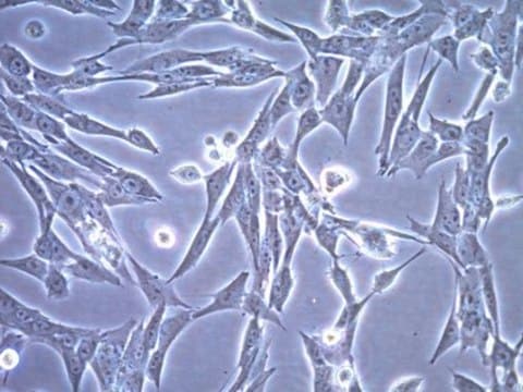
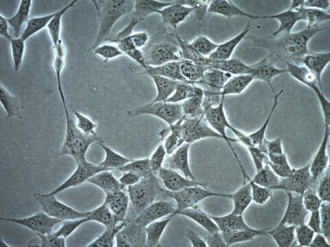
The CT-2A-Luc cell line is derived from a sub-cutaneous, non-metastatic murine glioma (astrocytoma) and is stably transduced with a firefly luciferase-EGFP reporter. CT-2A-Luc cells are marked by high levels of complex gangliosides and low distribution of the anti-angiogenic ganglioside GM3, as well as deficiency in the tumor suppressor PTEN/TSC2, a characteristic present in up to 70% of human high-grade glioma cell lines (2,3). CT-2A tumors are wild-type for p53 and recapitulate several features of human high-grade glioma, including high mitotic index and cell density, nuclear polymorphism, hemorrhage, pseudopalisading necrosis, and microvascular proliferation (4,5).

Source:
The parental CT-2A cell line was generated from a malignant astrocytoma formed via implantation of the carcinogen 20-methylcholanthrene in the cerebrum of a C57BL/6J mouse (6). The tumor was maintained through serial intracranial transplants prior to cell line isolation. The CT-2A-Luc luciferase cell line was clonally derived from CT-2A cells transduced with a lentiviral vector harboring a firefly luciferase (Fluc)-IRES-GFP cassette under control of the CMV promoter; cells were subsequently sorted for EGFP expression (7).

Cell Line Description

Cancer Cells

Application

Research Category
Cancer

Oncology
This product is intended for sale and sold solely to academic institutions for internal academic research use per the terms of the “Academic Use Agreement” as detailed in the product documentation. For information regarding any other use, please contact licensing@emdmillipore.com.

Quality

• Each vial contains 1X10⁶ viable cells.
• Cells are tested negative for infectious diseases by a Mouse Essential CLEAR panel by Charles River Animal Diagnostic Services.
• Cells are verified to be of mouse origin and negative for inter-species contamination from rat, chinese hamster, Golden Syrian hamster, human and non-human primate (NHP) as assessed by a Contamination CLEAR panel by Charles River Animal Diagnostic Services.
• Cells are negative for mycoplasma contamination

Storage and Stability

Store in liquid nitrogen. The cells can be cultured for at least 10 passages after initial thawing without significantly affecting the cell marker expression and functionality.

Storage Class Code

12 - Non Combustible Liquids

WGK

WGK 2

Flash Point(F)

does not flash

Flash Point(C)

does not flash


Certificates of Analysis (COA)

Search for Certificates of Analysis (COA) by entering the products Lot/Batch Number. Lot and Batch Numbers can be found on a product’s label following the words ‘Lot’ or ‘Batch’.

Already Own This Product?

Find documentation for the products that you have recently purchased in the Document Library.

Visit the Document Library

Michael Weller et al.
Neuro-oncology, 15(1), 4-27 (2012-11-09)
Newly diagnosed glioblastoma is now commonly treated with surgery, if feasible, or biopsy, followed by radiation plus concomitant and adjuvant temozolomide. The treatment of recurrent glioblastoma continues to be a moving target as new therapeutic principles enrich the standards of
R Martínez-Murillo et al.
Histology and histopathology, 22(12), 1309-1326 (2007-08-21)
Animal models of glial-derived neoplasms are needed to study the biological mechanisms of glioma tumorigenesis and those that sustain the disease state. With the aim to develop and characterize a suitable in vivo experimental mouse model for infiltrating astrocytoma, with
M Cotterchio et al.
Journal of lipid research, 35(1), 10-14 (1994-01-01)
The content of serum gangliosides was examined in VM and C57BL/6J (B6) mice that contained subcutaneous metastatic (VM) and non-metastatic (CT-2A) brain tumors, respectively. Gas-liquid chromatography (GLC) and high performance thin-layer chromatography (HPTLC) were used to analyze the serum gangliosides.
Emanuela Binello et al.
Journal of Cancer, 3, 166-174 (2012-04-20)
Evidence has pointed to brain tumor stem cells (BTSC) as culprits behind human high-grade glioma (hHGG) resistance to standard therapy. Pre-clinical rodent models are the mainstay for testing of new therapeutic strategies. The typical model involves the intracranial injection of

Our team of scientists has experience in all areas of research including Life Science, Material Science, Chemical Synthesis, Chromatography, Analytical and many others.

Contact Technical Service