Skip to Content
Merck
All Photos(13)

Key Documents

SAB4504210

Sigma-Aldrich

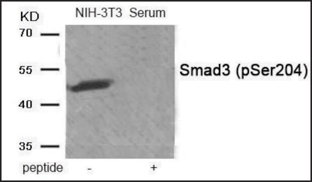
Anti-phospho-Smad3 (pSer204) antibody produced in rabbit

affinity isolated antibody

Sign Into View Organizational & Contract Pricing


About This Item

UNSPSC Code:
12352203
NACRES:
NA.41

biological source

rabbit

conjugate

unconjugated

antibody form

affinity isolated antibody

antibody product type

primary antibodies

clone

polyclonal

form

buffered aqueous solution

mol wt

antigen 48 kDa

species reactivity

mouse, human, rat

concentration

~1 mg/mL

technique(s)

ELISA: 1:20000
western blot: 1:500-1:1000

NCBI accession no.

UniProt accession no.

application(s)

research pathology

shipped in

wet ice

storage temp.

−20°C

target post-translational modification

phosphorylation (pSer204)

Gene Information

human ... SMAD3(4088)

General description

The SMAD3 (mothers against decapentaplegic homolog 3) gene is mapped to human chromosome 15q22.33. SMAD proteins contains three main domains : MH1 (MAD homology 1) - amino terminal domain (highly conserved), MH2 - carboxy terminal domain and a linker domain between the two domains. The MH1 domain of Smad3 serves as a DNA binding motif.(8)

Immunogen

The antiserum was produced against synthesized peptide derived from human Smad3 around the phosphorylation site of Ser204.

Immunogen Range: 170-219

Application

Anti-phospho-Smad3 (pSer204) antibody produced in rabbit has been used in western blot analysis.(7)

Biochem/physiol Actions

Smad3 (mothers against decapentaplegic homolog 3) protein identifies tandem repeats of the palindromic sequence GTCTAGAC, which is part of TGF-β (transforming growth factor β) signaling promoter region. Smad3 is associated with TGF-β (transforming growth factor beta) signaling pathway, a cellular process regulating the homeostasis, development, and repair of cartilage. It is known to induce extracellular matrix production. Smad proteins are responsible for the transduction of signals from cell surface to the nucleus. Phosphorylation of Smad3 controls tumor progression, inflammation, fibrosis, obesity and diabetes. Smad3 expression might be associated with the pathogenesis of osteoarthritis.

Features and Benefits

Evaluate our antibodies with complete peace of mind. If the antibody does not perform in your application, we will issue a full credit or replacement antibody. Learn more.

Physical form

Rabbit IgG in phosphate buffered saline (without Mg2+ and Ca2+), pH 7.4, 150mM NaCl, 0.02% sodium azide and 50% glycerol.

Disclaimer

Unless otherwise stated in our catalog or other company documentation accompanying the product(s), our products are intended for research use only and are not to be used for any other purpose, which includes but is not limited to, unauthorized commercial uses, in vitro diagnostic uses, ex vivo or in vivo therapeutic uses or any type of consumption or application to humans or animals.

Not finding the right product?  

Try our Product Selector Tool.

Storage Class Code

10 - Combustible liquids

WGK

WGK 1

Flash Point(F)

Not applicable

Flash Point(C)

Not applicable


Certificates of Analysis (COA)

Search for Certificates of Analysis (COA) by entering the products Lot/Batch Number. Lot and Batch Numbers can be found on a product’s label following the words ‘Lot’ or ‘Batch’.

Already Own This Product?

Find documentation for the products that you have recently purchased in the Document Library.

Visit the Document Library

Sodium tanshinone IIA sulfonate attenuates the transforming growth factor-beta1-induced differentiation of atrial fibroblasts into myofibroblasts in vitro
Yang L, et al.
International Journal of Molecular Medicine, 35(4), 1026-1032 (2015)
Down-regulation of microRNA-216b inhibits IL-1beta-induced chondrocyte injury by up-regulation of Smad3
He J, et al.
Bioscience Reports, 322(1), BSR20160588-BSR20160588 (2017)
Signaling via Smad2 and Smad3 is dispensable for adult murine hematopoietic stem cell function in vivo
Billing M, et al.
Experimental Hematology, 55(6), 34-44 (2017)
Cardiovascular phenotype in Smad3 deficient mice with renovascular hypertension
Kashyap S, et al.
PLoS ONE, 12(10), e0187062-e0187062 (2017)
Kristian Almstrup et al.
Cell and tissue research, 322(1), 159-165 (2005-04-23)
Testicular cancer is the most common malignancy among men in the reproductive age and the incidence is increasing, probably caused by environmental factors. Most testicular cancers are testicular germ cell tumours and all originate from a carcinoma in situ (CIS)

Our team of scientists has experience in all areas of research including Life Science, Material Science, Chemical Synthesis, Chromatography, Analytical and many others.

Contact Technical Service