Skip to Content
Merck
All Photos(1)

Key Documents

MABC1112

Sigma-Aldrich

Anti-Diasialoganglioside GD3 Antibody, clone R24

clone R24, from mouse

Synonym(s):

GD3, Ganglioside GD3, Disialoganglioside GD3

Sign Into View Organizational & Contract Pricing


About This Item

UNSPSC Code:
12352203
eCl@ss:
32160702

biological source

mouse

Quality Level

antibody form

purified antibody

antibody product type

primary antibodies

clone

R24, monoclonal

species reactivity

human, mouse, rat

technique(s)

affinity binding assay: suitable
flow cytometry: suitable
immunohistochemistry (formalin-fixed, paraffin-embedded sections): suitable
immunoprecipitation (IP): suitable
western blot: suitable

isotype

IgG3κ

shipped in

ambient

target post-translational modification

unmodified

Gene Information

human ... ST8SIA1(6489)

Related Categories

General description

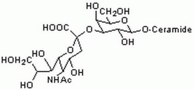
Gangliosides are sialic acid-containing oligoglycosylceramides. They are classified based on the number of sialic acid residues per molecule. GM gangliosides contain one sialic acid residue, GD gangliosides contain two, GT contain three, and GQ contain four sialic acid residues. Sialyltransferase-catalyzed 2-3 sialylation of the galactose of the trisaccharide Gal 1-4Glc 1-1 Cer produces GM3, which can be further sialylated to produce GD3 with two sialic acid on the galactose. GD3 is found in melanocytes and other cells that originate in the neuroectoderm. It is expressed almost exclusively among gangliosides in the early stage of the brain development of mice and rats. It is also detected in the T cell malignant leukocytes, such as acute lymphoblastic leukemia cells, adult T cell leukemia cells, and in activated normal human T lymphocytes. GD3 is found in almost all melanomas and is considered as an important biomarker for malignant melanoma. GD3 positive cells show markedly increased cell growth and invasive characteristics. Suppression of GD3 expression with antisense GD3 synthase cDNA is shown to reduce tumor cell phenotypes such as growth, migration, metastasis, and angiogenesis in F11 cells. Intra-cytoplasmic domain of neogenin is reported to act as a driver to promote various cancer phenotypes. Ref.: Hamamura, K et al. (2005). Proc. Natl. Acad. Sci. USA. 102 (31), 11041 11046; Dippold, WG et al. (1985). Cancer Res. 45(8): 3699-3705; Kaneko, K et al (2016). J. Biol. Chem. (In press).

Specificity

Clone R24 (a.k.a. R-24) immunostained neurites of cultured DRG neurons from wild-type, but not GD3 synthase-knockout, mice (Ribeiro-Resende, V.T., et al. (2014). PLoS One. 9(10):e108919).
Target glycolipid is not species-specific.

Immunogen

SK-MEL-28 human melanoma cells (Dippold, W.G., et al. (1980). Proc. Natl. Acad. Sci. U.S.A. 77(10):6114-6118).

Application

Affects Function: A representative lot induced cellular Lyn kinase activity and tyrosine phosphorylation of cellular proteins of primary rat cerebellar cultures in a time-dependent manner (Kasahara, K., et al. (1997). J. Biol. Chem. 272(47):29947-29953).

Affinity Binding Assay: A representative lot reacted with heat stable glycolipid on the surface of all SK-MEL melanoma and two of the five astrocytoma cells tested, whereas epithelial cell types, fibroblasts, and cells of hematopoietic origin were found to lack the surface antigen recognized by clone R24 (Dippold, W.G., et al. (1980). Proc. Natl. Acad. Sci. U.S.A. 77(10):6114-6118).

Flow Cytometry Analysis: A representative lot immunostained the surface of cultured primary rat cerebellar cells (Kasahara, K., et al. (1997). J. Biol. Chem. 272(47):29947-29953).

Immunocytochemistry Analysis: A representative lot immunostained NGF-induced nurite outgrowths of cultured DRG neurons from wild-type, but not GD3 synthase-knockout, mice (Ribeiro-Resende, V.T., et al. (2014). PLoS One. 9(10):e108919).

Immunohistochemistry Analysis: A representative lot detected GD3 immunoreactivity in unfixed frozen tissue samples of primary melanoma and metastatic malignant melanoma (Dippold, W.G., et al. (1985). Cancer Res. 45(8):3699-3705).

Immunoprecipitation Analysis: A representative lot co-immunoprecipitated Lyn from rat brain membrane extract. Clone R24 co-immunoprecipitated caveolin and the exogenously expressed Lyn, but not Src, from CHO cells expressing exogenous GD3 synthase (Kasahara, K., et al. (1997). J. Biol. Chem. 272(47):29947-29953).
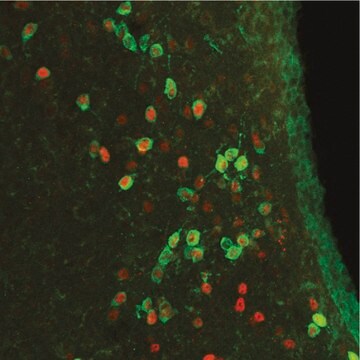
Detect ganglioside GD3 using this mouse monoclonal Anti-Diasialoganglioside GD3, clone R24 Antibody, Cat. No. MABC1112, validated for use in Flow Cytometry and Immunocytochemistry, Western Blotting, Affects Function, Immunoprecipitation, Immunohistochemistry (Paraffin), Immunohistochemistry, Affinit
Research Category
Apoptosis & Cancer

Quality

Evaluated by Flow Cytometry Analysis in SK-MEL-28 human skin melanoma cells.

Flow Cytometry Analysis: 0.2 µL of this antibody detected GD3 immunoreactivity on the surface of one million SK-MEL-28 human skin melanoma cells.

Physical form

Format: Purified
Protein G purified
Purified mouse monoclonal antibody IgG3 in PBS without azide.

Storage and Stability

Stable for 1 year at -20°C from date of receipt.

Handling Recommendations: Upon receipt and prior to removing the cap, centrifuge the vial and gently mix the solution. Aliquot into microcentrifuge tubes and store at -20°C. Avoid repeated freeze/thaw cycles, which may damage IgG and affect product performance.

Other Notes

Concentration: Please refer to lot specific datasheet.

Disclaimer

Unless otherwise stated in our catalog or other company documentation accompanying the product(s), our products are intended for research use only and are not to be used for any other purpose, which includes but is not limited to, unauthorized commercial uses, in vitro diagnostic uses, ex vivo or in vivo therapeutic uses or any type of consumption or application to humans or animals.

Not finding the right product?  

Try our Product Selector Tool.

Storage Class Code

12 - Non Combustible Liquids

WGK

WGK 2

Flash Point(F)

Not applicable

Flash Point(C)

Not applicable


Certificates of Analysis (COA)

Search for Certificates of Analysis (COA) by entering the products Lot/Batch Number. Lot and Batch Numbers can be found on a product’s label following the words ‘Lot’ or ‘Batch’.

Already Own This Product?

Find documentation for the products that you have recently purchased in the Document Library.

Visit the Document Library

Our team of scientists has experience in all areas of research including Life Science, Material Science, Chemical Synthesis, Chromatography, Analytical and many others.

Contact Technical Service