Skip to Content
Merck
All Photos(3)

Key Documents

AB756P

Sigma-Aldrich

Anti-Collagen Antibody, Type IV

Chemicon®, from rabbit

Synonym(s):

Anti-ATS2, Anti-CA44

Sign Into View Organizational & Contract Pricing


About This Item

UNSPSC Code:
12352203
eCl@ss:
32160702
NACRES:
NA.41

biological source

rabbit

Quality Level

antibody form

purified antibody

antibody product type

primary antibodies

clone

polyclonal

species reactivity

mouse

manufacturer/tradename

Chemicon®

technique(s)

ELISA: suitable
immunofluorescence: suitable
immunohistochemistry: suitable (paraffin)

suitability

not suitable for Western blot

UniProt accession no.

shipped in

dry ice

target post-translational modification

unmodified

General description

Collagen Type IV extracted and purified from mouse tumor tissues. Antibody shows less than 0.1% reactivity with human Collagen types IV, V and mouse Collagen Types I, II, III and mouse Fibronectin and Laminin.
Collagen is the main protein of connective tissue in animals and the most abundant protein in mammals, making up about 25% of the total protein content. It is one of the long, fibrous structural proteins whose functions are quite different from those of globular proteins such as enzymes; tough bundles of collagen called collagen fibers are a major component of the extracellular matrix that supports most tissues and gives cells structure from the outside, but collagen is also found inside certain cells. Collagen has great tensile strength, and is the main component of fascia, cartilage, ligaments, tendons, bone and teeth. Along with soft keratin, it is responsible for skin strength and elasticity, and its degradation leads to wrinkles that accompany aging. It strengthens blood vessels and plays a role in tissue development. It is present in the cornea and lens of the eye in crystalline form. Collagen occurs in many places throughout the body. There are 12 types of collagen described in literature. Collagen Type 1: This is the most abundant collagen of the human body. It is present in scar tissue, the end product when tissue heals by repair. It is found in tendons, the endomysium of myofibrils and the organic part of bone. Collagen Type 4: It is present in basal lamina and the eye lens. Also serves as part of the filtration system in capillaries and the glomeruli of nephron in the kidney.

Specificity

Antibody shows less than 0.1% reactivity with human Collagen types IV, V and mouse Collagen Types I, II, III, mouse Fibronectin, and mouse laminin.
Mouse Collagen, Type IV: 100 (%’s at a 1:5000 RIA dilution) Mouse Collagen, Types I, II, III: <0.1 Human Collagen, Types IV, V: <0.1 Mouse Fibronectin: <0.1 Mouse Laminin: <0.1 Reactivity with other species has not been determined.

Immunogen

Collagen Type IV extracted and purified from mouse tumor tissues.

Application

ELISA:
A previous lot of this antibody was used in ELISA at >1:200 (OD >500).

Immunofluorescence:
A previous lot of this antibody was used in immunofluorescence.

Immunohistochemistry:
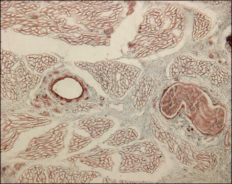
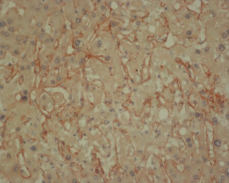
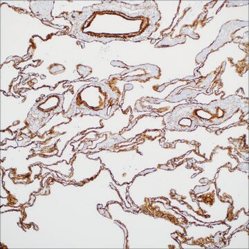
1:80 dilution for immunofluorescent staining of fresh frozen mouse skin and liver tissues. Acetone or methyl-carnoy fixed paraffin-embedded tissue (mouse skin and liver) is also reactive.

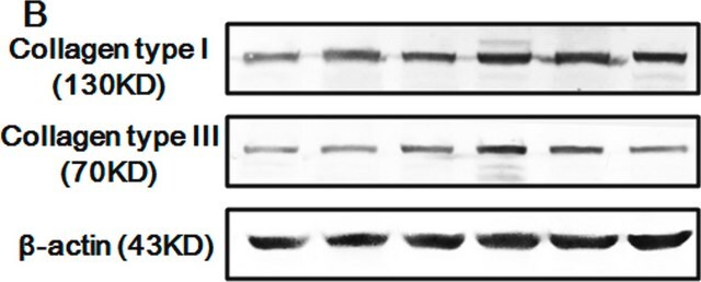
Not recommended for Western blots.

Optimal working dilutions must be determined by end user.
Research Category
Cell Structure
Research Sub Category
ECM Proteins
This Anti-Collagen Antibody, Type IV is validated for use in ELISA, IF, IH(P) for the detection of Collagen.

Target description

139 kDa (unprocessed precursor)

Linkage

Replaces: AB756

Physical form

Format: Purified
Protein G Purified
The purified antibody is supplied in a buffer containing a mixture of 0.1M Citrate, 0.1M potassium phosphate, at a pH of 7.2-7.4 and 10µl/ml of antibiotics and antimycotics.

Storage and Stability

Stable for 1 year at -20ºC from date of receipt.

Analysis Note

Control
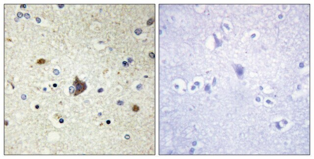
Positive Control: Kidney, muscle, tendon spleen tissue, mouse liver. Negative Control: Neurons/glia.

Other Notes

Concentration: Please refer to the Certificate of Analysis for the lot-specific concentration.

Legal Information

CHEMICON is a registered trademark of Merck KGaA, Darmstadt, Germany

Disclaimer

Unless otherwise stated in our catalog or other company documentation accompanying the product(s), our products are intended for research use only and are not to be used for any other purpose, which includes but is not limited to, unauthorized commercial uses, in vitro diagnostic uses, ex vivo or in vivo therapeutic uses or any type of consumption or application to humans or animals.

Not finding the right product?  

Try our Product Selector Tool.

Storage Class Code

12 - Non Combustible Liquids

WGK

WGK 1

Flash Point(F)

Not applicable

Flash Point(C)

Not applicable


Certificates of Analysis (COA)

Search for Certificates of Analysis (COA) by entering the products Lot/Batch Number. Lot and Batch Numbers can be found on a product’s label following the words ‘Lot’ or ‘Batch’.

Already Own This Product?

Find documentation for the products that you have recently purchased in the Document Library.

Visit the Document Library

Retinal vascular development is mediated by endothelial filopodia, a preexisting astrocytic template and specific R-cadherin adhesion.
Michael I Dorrell, Edith Aguilar, Martin Friedlander
Investigative Ophthalmology & Visual Science null
Long-term retinal PEDF overexpression prevents neovascularization in a murine adult model of retinopathy.
Haurigot, V; Villacampa, P; Ribera, A; Bosch, A; Ramos, D; Ruberte, J; Bosch, F
Testing null
Retinal O-linked N-acetylglucosamine protein modifications: implications for postnatal retinal vascularization and the pathogenesis of diabetic retinopathy.
Gurel, Zafer, et al.
Molecular Vision, 19, 1047-1059 (2013)
Sonic hedgehog-dependent synthesis of laminin alpha1 controls basement membrane assembly in the myotome.
Anderson, C; Thorsteinsdottir, S; Borycki, AG
Development null
Breaches of the pial basement membrane and disappearance of the glia limitans during development underlie the cortical lamination defect in the mouse model of muscle-eye-brain disease.
Huaiyu Hu,Yuan Yang,Amber Eade,Yufang Xiong,Yue Qi
The Journal of Comparative Neurology null

Our team of scientists has experience in all areas of research including Life Science, Material Science, Chemical Synthesis, Chromatography, Analytical and many others.

Contact Technical Service