1.04302
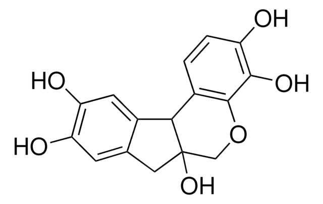
Hematoxylin cryst. (C.I. 75290)
for microscopy
Synonym(s):
Hematoxylin cryst. (C.I. 75290)
About This Item
Recommended Products
Quality Level
form
solid
IVD
for in vitro diagnostic use
technique(s)
microbe id | staining: suitable
visual transition interval
5.0-7.2, yellow to violet
mp
140 °C (Elimination of water of crystallisation)
bulk density
500 kg/m3
application(s)
diagnostic assay manufacturing
hematology
histology
storage temp.
2-30°C
SMILES string
O1C[C@@]2([C@H](c4c1c(c(cc4)O)O)c3c(cc(c(c3)O)O)C2)O
InChI
1S/C16H14O6/c17-10-2-1-8-13-9-4-12(19)11(18)3-7(9)5-16(13,21)6-22-15(8)14(10)20/h1-4,13,17-21H,5-6H2/t13-,16+/m1/s1
InChI key
WZUVPPKBWHMQCE-CJNGLKHVSA-N
Looking for similar products? Visit Product Comparison Guide
General description
It is an IVD registered product and CE certified, thus can be used for clinical diagnostic purposes. For more details, please see instructions for use (IFU).
Signal Word
Warning
Hazard Statements
Precautionary Statements
Hazard Classifications
Eye Irrit. 2
Storage Class Code
11 - Combustible Solids
WGK
WGK 1
Flash Point(F)
Not applicable
Flash Point(C)
Not applicable
Certificates of Analysis (COA)
Search for Certificates of Analysis (COA) by entering the products Lot/Batch Number. Lot and Batch Numbers can be found on a product’s label following the words ‘Lot’ or ‘Batch’.
Already Own This Product?
Find documentation for the products that you have recently purchased in the Document Library.
Customers Also Viewed
Our team of scientists has experience in all areas of research including Life Science, Material Science, Chemical Synthesis, Chromatography, Analytical and many others.
Contact Technical Service