Skip to Content
Merck
All Photos(1)

Documents

C7048

Sigma-Aldrich

Monoclonal Anti-CD3 antibody produced in mouse

clone UCHT-1, purified immunoglobulin, buffered aqueous solution

Synonym(s):

Monoclonal Anti-CD3

Sign Into View Organizational & Contract Pricing


About This Item

UNSPSC Code:
12352203
NACRES:
NA.41

biological source

mouse

Quality Level

conjugate

unconjugated

antibody form

purified immunoglobulin

antibody product type

primary antibodies

clone

UCHT-1, monoclonal

form

buffered aqueous solution

species reactivity

human

technique(s)

flow cytometry: 5 μL using 1 × 10⁶ cells

isotype

IgG1

UniProt accession no.

shipped in

wet ice

storage temp.

2-8°C

target post-translational modification

unmodified

Gene Information

human ... CD3E(916)

Looking for similar products? Visit Product Comparison Guide

Related Categories

Specificity

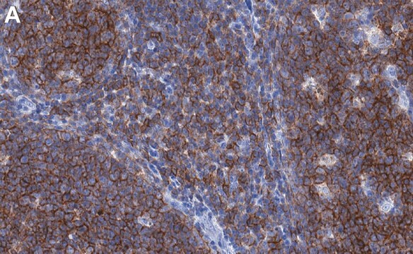
Recognizes the CD3 complex. The epitope recognized by clone no. UCHT-1 is expressed on the ε-chain of the CD3 antigen/T cell receptor complex. 3rd Workshop: code no. 126, 208 and 471
4th Workshop: paper no. T3.2

Immunogen

human thymocytes followed by Sezary T cells.

Application

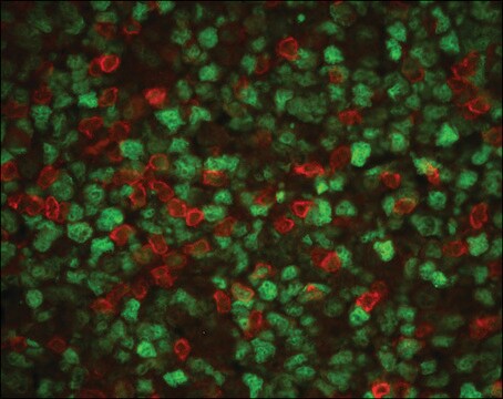
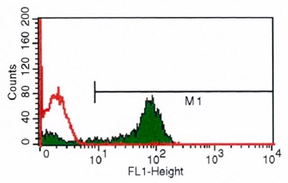
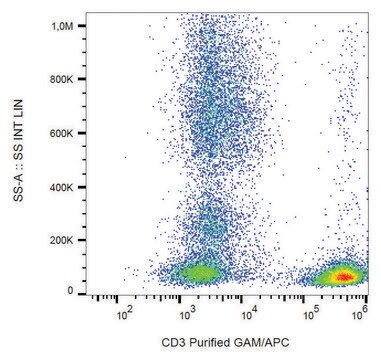
Monoclonal Anti-CD3 antibody produced in mouse is suitable for flow cytometry at a dilution of 5μL using 1 × 10⁶ cells.

Biochem/physiol Actions

The CD3e cytoplasmic domain of the T cell receptor binds to the inner leaflet of the plasma membrane (PM) and is accessible during early stages of T cell activation. It is specific for T lymphocytes, found in Purkinje cells of the cerebellum and gastric parietal cells of humans and other species. The antibodies commonly used to recognize CD3 are directed against the ε-subunit of the T cell receptor. Cytoplasmic cyCD3 expression can only be achieved during the early phase of NK-cell differentiation.

Target description

The CD3 complex (16/20/29/22/25-28 kDa) consists of 5 polypeptide chains associated with each other and with the T cell receptor.

Physical form

Solution in 0.01 M phosphate buffered saline, pH 7.4, containing 1% bovine serum albumin and 15 mM sodium azide.

Disclaimer

Unless otherwise stated in our catalog or other company documentation accompanying the product(s), our products are intended for research use only and are not to be used for any other purpose, which includes but is not limited to, unauthorized commercial uses, in vitro diagnostic uses, ex vivo or in vivo therapeutic uses or any type of consumption or application to humans or animals.

Not finding the right product?  

Try our Product Selector Tool.

Storage Class Code

10 - Combustible liquids

WGK

nwg

Flash Point(F)

Not applicable

Flash Point(C)

Not applicable


Certificates of Analysis (COA)

Search for Certificates of Analysis (COA) by entering the products Lot/Batch Number. Lot and Batch Numbers can be found on a product’s label following the words ‘Lot’ or ‘Batch’.

Already Own This Product?

Find documentation for the products that you have recently purchased in the Document Library.

Visit the Document Library

Francesco Dieli et al.
The Journal of infectious diseases, 186(12), 1835-1839 (2002-11-26)
Vgamma9/Vdelta2 T cells can contribute to protective immune response against Mycobacterium tuberculosis, although the extent to which and mechanisms by which they could actually protect against human tuberculosis remain unclear. We have previously reported that Vgamma9/Vdelta2 T cells from tuberculin
Magda De Smedt et al.
Blood, 110(7), 2696-2703 (2007-07-17)
It has been proposed that heterogeneity in natural killer (NK)-cell phenotype and function can be achieved through distinct thymic and bone marrow pathways of NK-cell development. Here, we show a link between Notch signaling and the generation of intracellular CD3epsilon
Joseph Alroy et al.
Pathology, research and practice, 201(12), 803-808 (2005-11-26)
CD3 antigen, formerly thought to be specific for T lymphocytes, has been identified in Purkinje cells of the cerebellum and gastric parietal cells in several species, including humans. The antibodies commonly used to recognize CD3 are directed against the epsilon-subunit
Luciën E P M van der Vlugt et al.
The Journal of infectious diseases, 210(8), 1207-1216 (2014-05-06)
Chronic schistosome infections are associated with T-cell hyporesponsiveness and a strong regulatory network. Murine studies have shown that schistosome infections can induce regulatory CD1d(hi) B cells, which inhibit inflammatory responses. Here, we evaluated the influence of regulatory B cells (Bregs)
Rinat Sharir et al.
Cardiovascular research, 103(4), 585-596 (2014-06-27)
Ischaemic damage is associated with up-regulation of pro-inflammatory cytokines, as well as invasion of leucocytes and lymphocytes to the injured muscle. Regulatory T cells (Tregs) exert suppressive effects on several immune and non-immune cellular elements. We hypothesized that adoptive Treg

Our team of scientists has experience in all areas of research including Life Science, Material Science, Chemical Synthesis, Chromatography, Analytical and many others.

Contact Technical Service