Z764000
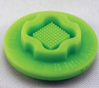
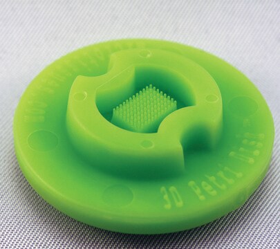
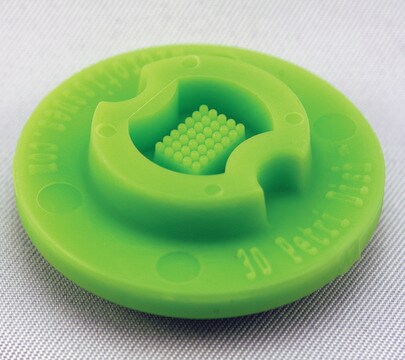
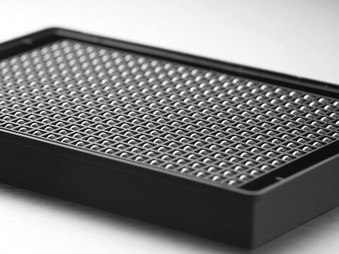
MicroTissues® 3D Petri Dish® micro-mold spheroids
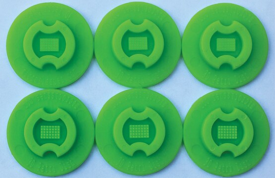
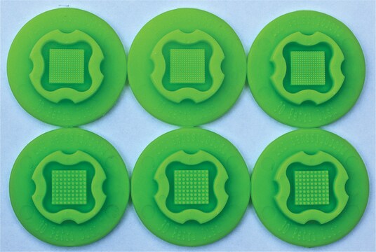
size S, 16 x 16 array, fits 12 well plates
Synonyme(s) :
3D, 3D Cell Culture
About This Item
Produits recommandés
Matériaux
spherical
Taille
S
Stérilité
sterile; autoclaved
Caractéristiques
lid: no
16 x 16 array
Conditionnement
pack of 6 ea
Fabricant/nom de marque
MicroTissues Inc. 12-256
Volume
190 μL
Vous recherchez des produits similaires ? Visite Guide de comparaison des produits
Description générale
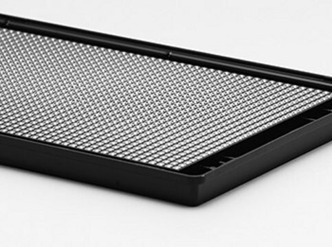
- Nominal dimensions of each 3D culture recess: diam. 300 μm x D 800 μm
- Micro-molds also form single chamber for cell seeding
Since the agarose is transparent, the spheroids or microtissues that form at the bottom of each agarose micro-well can be easily viewed using a standard inverted microscope using phase contrast, bright field or fluorescence microscopy.
- The micro-molds are reusable up to 12 times and can be sterilized via a standard steam autoclave (30 min, dry cycle)
- The micro-molds should be stored in a covered container to maintain sterility and to avoid collecting dust or fibres on their small features
- Any microscope with sufficient magnification and resolution functions for cell based views or images will be suitable for use with the 3D Petri Dish products
Informations légales
Faites votre choix parmi les versions les plus récentes :
Certificats d'analyse (COA)
It looks like we've run into a problem, but you can still download Certificates of Analysis from our Documents section.
Si vous avez besoin d'assistance, veuillez contacter Service Clients
Déjà en possession de ce produit ?
Retrouvez la documentation relative aux produits que vous avez récemment achetés dans la Bibliothèque de documents.
Les clients ont également consulté
Notre équipe de scientifiques dispose d'une expérience dans tous les secteurs de la recherche, notamment en sciences de la vie, science des matériaux, synthèse chimique, chromatographie, analyse et dans de nombreux autres domaines..
Contacter notre Service technique
