Accéder au contenu
Merck
Toutes les photos(1)

Principaux documents

P4110

Sigma-Aldrich

Monoclonal Anti-Phosphotyrosine antibody produced in mouse

clone PY20, purified immunoglobulin, buffered aqueous glycerol solution

Synonyme(s) :

Monoclonal Anti-Phosphotyrosine, Phospho-Tyr, Phospho-tyrosine, p-Tyr

Se connecterpour consulter vos tarifs contractuels et ceux de votre entreprise/organisme


About This Item

Code UNSPSC :
12352203
Nomenclature NACRES :
NA.44

Source biologique

mouse

Niveau de qualité

Conjugué

unconjugated

Forme d'anticorps

purified immunoglobulin

Type de produit anticorps

primary antibodies

Clone

PY20, monoclonal

Forme

buffered aqueous glycerol solution

Technique(s)

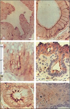
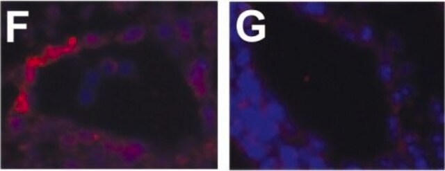
immunohistochemistry: suitable
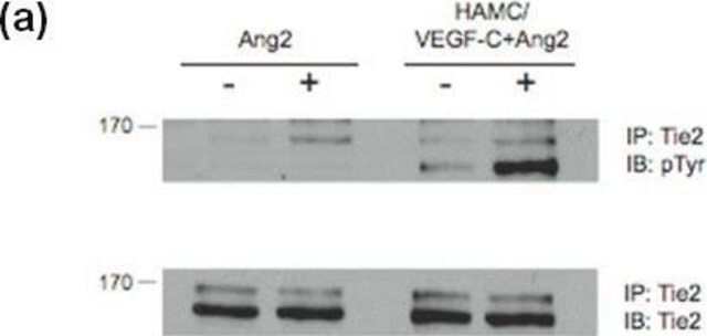
immunoprecipitation (IP): suitable
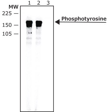
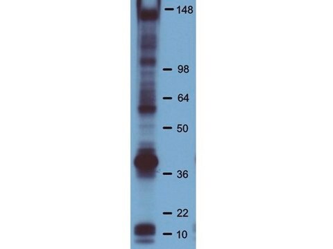
western blot: suitable

Isotype

IgG2b

Conditions d'expédition

wet ice

Température de stockage

−20°C

Modification post-traductionnelle de la cible

phosphorylation (pTyr)

Vous recherchez des produits similaires ? Visite Guide de comparaison des produits

Description générale

Mouse monoclonal clone PY20 anti-Phosphotyrosine is specific for both native and denatured proteins containing phosphorylated tyrosine. Antibody binding is inhibited with phosphotyrosine and phenylphosphate, but not phosphoeserine or phosphothreonine.
Protein phosphorylation is a basic mechanism for the modification of protein function in eukaryotic cells. Tyrosine phosphorylation is a rare post-translational event in normal tissue, accounting for only 0.03% of phosphorylated amino acids. The level of phosphorylated tyrosine in many cellular proteins increases tenfold following various activation processes that are mediated through phosphotyrosine kinases. The importance of tyrosine phosphorylation has been established by the demonstration that it is an integral response in many different mitogenic receptor systems.
Reversible phosphorylation of proteins is an important post-translational modification that plays a regulatory role in the expression of most proteins in the cells. Reversible phosphorylation at multiple serine, tyrosine and threonine residues mediate numerous signalling pathways in both prokaryotic and Eukaryotic cells. Cellular proteins with phosphorylated tyrosine increase many fold by the activation of tyrosine kinases. Most mitogenic receptor systems such as EGF, PDGF, insulin receptors contain tyrosine kinase domains that undergo autophosphorylation when receptors bind to the respective ligands. Tyrosine-specific protein kinase activity has also been described in many retroviral oncogene proteins. Cells transformed by these oncogenes contain elevated levels of phosphotyrosine. Many of the oncogenes found in mammalian oncogenic viruses encode tyrosine protein kinases that reside in the cellular cytoplasm. Others encode transmembrane receptors whose tyrosine phosphotransferase activity is stimulated by the binding of ligand to the extracellular domain.
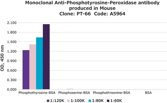
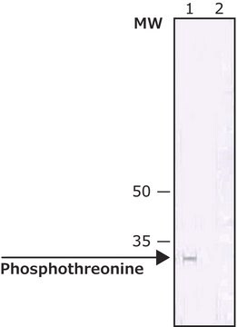
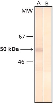
Monoclonal Anti-Phosphotyrosine is specific for both native and denatured proteins containing phosphorylated tyrosine.

Immunogène

phosphotyrosine-protein conjugate

Application

Monoclonal anti-phosphotyrosine antibody produced in mouse has been used in the detection of tyrosine phosphorylation in proteins by 2D western blotting and western blotting.

Forme physique

Solution in 20 mM phosphate buffered saline, pH 7.5, containing 50% glycerol and 3 mM sodium azide

Clause de non-responsabilité

Unless otherwise stated in our catalog or other company documentation accompanying the product(s), our products are intended for research use only and are not to be used for any other purpose, which includes but is not limited to, unauthorized commercial uses, in vitro diagnostic uses, ex vivo or in vivo therapeutic uses or any type of consumption or application to humans or animals.

Vous ne trouvez pas le bon produit ?  

Essayez notre Outil de sélection de produits.

Code de la classe de stockage

12 - Non Combustible Liquids

Classe de danger pour l'eau (WGK)

nwg

Point d'éclair (°F)

Not applicable

Point d'éclair (°C)

Not applicable


Faites votre choix parmi les versions les plus récentes :

Certificats d'analyse (COA)

Lot/Batch Number

Vous ne trouvez pas la bonne version ?

Si vous avez besoin d'une version particulière, vous pouvez rechercher un certificat spécifique par le numéro de lot.

Déjà en possession de ce produit ?

Retrouvez la documentation relative aux produits que vous avez récemment achetés dans la Bibliothèque de documents.

Consulter la Bibliothèque de documents

Huanghui Tang et al.
Biology of reproduction, 88(4), 96-96 (2013-03-08)
By targeted disruption of the lactate dehydrogenase c (Ldhc) gene, we demonstrated that spermatozoa require Ldhc for capacitation, motility, and fertilizing capacity. Ldhc expression is restricted to the developing germ cells that, however, are apparently not compromised by the lack
Human lactate dehydrogenase A (LDHA) rescues mouse Ldhc-null sperm function
Tang H, et al.
Biology of Reproduction, 88(4), 96-91 (2013)
Cellular response of human neuroblastoma cells to alpha-synuclein fibrils, the main constituent of Lewy bodies
Pieri L, et al.
Biochim. Biophys. Acta Gen. Subj., 1860(1), 8-19 (2016)
Toshiaki Fukushima et al.
Endocrinology, 149(7), 3729-3742 (2008-04-12)
Bioactivities of IGFs in various cells are often potentiated in the presence of other hormones. In previous studies we showed that pretreatment of rat FRTL-5 thyroid cells with TSH or other cAMP-generating agents markedly potentiated DNA synthesis induced by IGF-I.
Jixian Luo et al.
Frontiers in oncology, 10, 1512-1512 (2020-09-10)
Although we currently have a good understanding of the role C-X-C chemokine receptor type 4 (CXCR4) plays in T cell acute lymphoblastic leukemia (T-ALL), the mechanism of CXCR4-mediated T-ALL migration remains elusive. Therefore, we focus on the downstream signals of

Notre équipe de scientifiques dispose d'une expérience dans tous les secteurs de la recherche, notamment en sciences de la vie, science des matériaux, synthèse chimique, chromatographie, analyse et dans de nombreux autres domaines..

Contacter notre Service technique