Accéder au contenu
Merck
Toutes les photos(1)

Principaux documents

A6229

Sigma-Aldrich

Anti-AKR1C3 antibody, Mouse monoclonal

clone NP6.G6.A6, purified from hybridoma cell culture

Synonyme(s) :

Anti-DD-3, Anti-DD3, Anti-HA1753

Se connecterpour consulter vos tarifs contractuels et ceux de votre entreprise/organisme


About This Item

Numéro MDL:
Code UNSPSC :
12352203
Nomenclature NACRES :
NA.41

Source biologique

mouse

Niveau de qualité

Conjugué

unconjugated

Forme d'anticorps

purified immunoglobulin

Type de produit anticorps

primary antibodies

Clone

NP6.G6.A6, monoclonal

Forme

buffered aqueous solution

Poids mol.

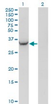
antigen ~38 kDa

Espèces réactives

human

Conditionnement

antibody small pack of 25 μL

Technique(s)

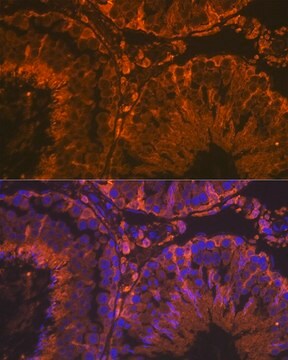
immunohistochemistry: suitable
indirect ELISA: suitable
microarray: suitable
western blot: 0.25-0.5 μg/mL using cytosolic fraction extract of A549 human lung carcinoma cell

Isotype

IgG1

Numéro d'accès UniProt

Conditions d'expédition

dry ice

Température de stockage

−20°C

Modification post-traductionnelle de la cible

unmodified

Informations sur le gène

human ... AKR1C3(8644)

Description générale

Anti-AKR1C3 antibody, Mouse monoclonal (humanα - hydroxysteroid dehydrogenase, type 2,3; 17β- hydroxysteroid dehydrogenase, type 5) (mouse IgG1 isotype) is derived from the hybridoma NP6.G6. A6 produced by the fusion of mouse myeloma cells (SP20 cells) and splenocytes from BALB/c mice immunized with human AKR1C3 protein. Oxidoreduction of natural and foreign substrates is performed by three enzymes superfamilies, one of them being the AKRs (aldo-keto reductases) family. This family contains 114 proteins that are expressed in prokaryotes and eukaryotes and is divided into 14 subfamilies (AKR1-AKR14). The AKR1 family is the largest among the different fourteen families and contains the aldose reductases, aldehyde reductases, hydroxysteroid dehydrogenases, and steroid 5 β-reductases. AKR1Cs are expressed in many tissues.

Immunogène

human AKR1C3 protein.

Application

Anti-AKR1C3 antibody, Mouse monoclonal has been used in
  • western blotting
  • immunohistochemistry
  • enzyme linked immunoassay (ELISA).

Applications in which this antibody has been used successfully, and the associated peer-reviewed papers, are given below.
Immunohistochemistry (1 paper)
Immunohistochemistry on human placentas were performed using monoclonal mouse anti-AKR1C3 at a dilution of 1:100 or western blot at 1:500.
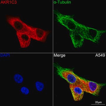
Mouse monoclonal clone NP6.G6.A6 anti-AKR1C3 antibody used to tag aldo-keto reductase 1C3 for detection and quantitation by immunocytochemical and immunohistochemical (IHC) techniques. It is used as a probe to determine the presence and roles of aldo-keto reductase 1C3 in the regulation of the androgen 5α-dihydrotestosterone (DHT) and 3α-diol compound levels in vivo.

Actions biochimiques/physiologiques

In humans AKR1C3 (aldo-keto reductase family 1 member C3) including AKR1C1,2 and 4, catalyze the reduction of the androgen 5α-dihydrotestosterone (DHT) into inactive 3β or 3α-androstane diol (3α/β-Diol). AKR1C1-4 isoforms also display 3α[17β]-hydroxysteroid oxidase activity using 3α-Diol as a substrate, in vitro. In non-small cell lung carcinoma, AKR1Cs expression is dramatically increased. Due to their product profile and discrete tissue localization, they may regulate the level of active androgens, estrogens, and progestins in target tissues.

Description de la cible

AKR1C3, aldo-keto reductase family 1, member C3 (3-α hydroxysteroid dehydrogenase, type II), encodes a member of the aldo/keto reductase superfamily, which consists of more than 40 known enzymes and proteins. These enzymes catalyze the conversion of

Forme physique

Solution in 0.01 M phosphate buffered saline, pH 7.4, containing 15 mM sodium azide.

Clause de non-responsabilité

Unless otherwise stated in our catalog or other company documentation accompanying the product(s), our products are intended for research use only and are not to be used for any other purpose, which includes but is not limited to, unauthorized commercial uses, in vitro diagnostic uses, ex vivo or in vivo therapeutic uses or any type of consumption or application to humans or animals.

Vous ne trouvez pas le bon produit ?  

Essayez notre Outil de sélection de produits.

Code de la classe de stockage

12 - Non Combustible Liquids

Classe de danger pour l'eau (WGK)

WGK 2

Point d'éclair (°F)

Not applicable

Point d'éclair (°C)

Not applicable

Équipement de protection individuelle

Eyeshields, Gloves, multi-purpose combination respirator cartridge (US)


Faites votre choix parmi les versions les plus récentes :

Certificats d'analyse (COA)

Lot/Batch Number

Vous ne trouvez pas la bonne version ?

Si vous avez besoin d'une version particulière, vous pouvez rechercher un certificat spécifique par le numéro de lot.

Déjà en possession de ce produit ?

Retrouvez la documentation relative aux produits que vous avez récemment achetés dans la Bibliothèque de documents.

Consulter la Bibliothèque de documents

Human cytosolic 3alpha-hydroxysteroid dehydrogenases of the aldo-keto reductase superfamily display significant 3beta-hydroxysteroid dehydrogenase activity: implications for steroid hormone metabolism and action
Steckelbroeck S, et al.
The Journal of Biological Chemistry, 279(11), 10784-10795 (2004)
Rob D Catalano et al.
Molecular human reproduction, 17(3), 182-192 (2010-11-30)
Prostanoids are well-described primary mediators of inflammatory processes and are essential for the normal physiological function of the female reproductive system. The aim of this study was to determine the temporal expression of the prostanoid biosynthetic enzymes (PTGS1, PTGS2, PTGES
Human 3alpha-hydroxysteroid dehydrogenase isoforms (AKR1C1-AKR1C4) of the aldo-keto reductase superfamily: functional plasticity and tissue distribution reveals roles in the inactivation and formation of male and female sex hormones
Penning TM, et al.
The Biochemical Journal, 351(27), 1-1 (2000)
The aldo-keto reductase superfamily homepage
Hyndman D, et al.
Chemico-Biological Interactions, 143-144(27), 621-631 (2003)
Julie J Loiselle et al.
Journal of environmental radioactivity, 196, 64-81 (2018-11-06)
Radon is the second leading cause of lung cancer, after tobacco smoke. While tobacco smoke-induced carcinogenesis has been studied extensively, far less is known about radon-induced carcinogenesis, particularly in relation to the influence of radon on gene expression. The objectives

Notre équipe de scientifiques dispose d'une expérience dans tous les secteurs de la recherche, notamment en sciences de la vie, science des matériaux, synthèse chimique, chromatographie, analyse et dans de nombreux autres domaines..

Contacter notre Service technique